|
|
|
|
|
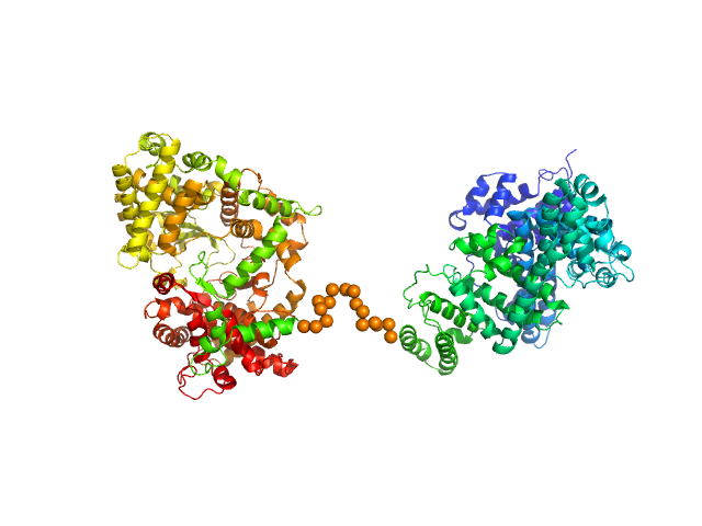
| Sample: |
Collagen like-peptide [GPRG(POG)13] trimer, 11 kDa protein
Collagenase ColH (Polycystic kidney disease domain 2 (PKD2) and Collagen binding domain (CBD) with Tyr780Ser, His782Ser, Tyr796Ser and Tyr801Ser) monomer, 23 kDa Hathewaya histolytica protein
|
| Buffer: |
50 mM HEPES, 100 mM NaCl, 5 mM CaCl2, pH: 7.5 |
| Experiment: |
SAXS
data collected at 12.3.1 (SIBYLS), Advanced Light Source (ALS) on 2019 Mar 5
|
Elucidating Collagen Degradation Synergy between Col G and Col H from Hathewaya (Clostridium) histolytica and Identifying novel structural features in HPT and REC domains from VarS histidine kinase in V. alginolyticus
University of Arkansas PhD thesis 28030553 (2020)
Perry Caviness
|
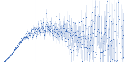
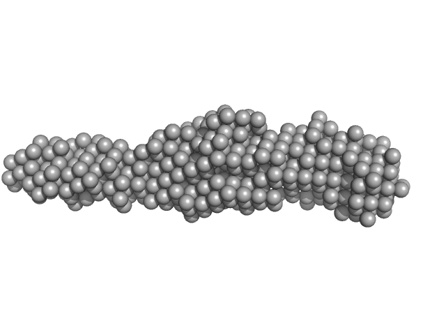
| RgGuinier |
3.2 |
nm |
| Dmax |
18.0 |
nm |
| VolumePorod |
34 |
nm3 |
|
|
|
|
|
|
|
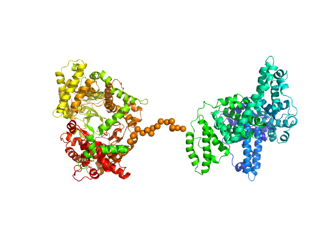
| Sample: |
Collagenase ColH (Polycystic kidney disease domain 2 (PKD2) and Collagen binding domain (CBD)) monomer, 23 kDa Hathewaya histolytica protein
Collagen like-peptide [GPRG(POG)13] trimer, 11 kDa protein
|
| Buffer: |
50 mM HEPES, 100 mM NaCl, 5 mM CaCl2, pH: 7.5 |
| Experiment: |
SAXS
data collected at 12.3.1 (SIBYLS), Advanced Light Source (ALS) on 2019 Mar 5
|
Elucidating Collagen Degradation Synergy between Col G and Col H from Hathewaya (Clostridium) histolytica and Identifying novel structural features in HPT and REC domains from VarS histidine kinase in V. alginolyticus
University of Arkansas PhD thesis 28030553 (2020)
Perry Caviness
|
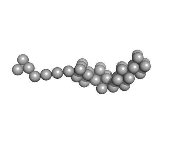
| RgGuinier |
3.3 |
nm |
| Dmax |
18.0 |
nm |
| VolumePorod |
35 |
nm3 |
|
|
|
|
|
|
|
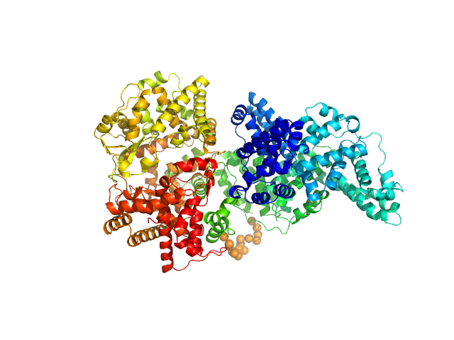
| Sample: |
Cell wall synthesis protein Wag31 tetramer, 34 kDa Mycobacterium tuberculosis protein
|
| Buffer: |
20 mM Tris pH 7.5, 150 mM NaCl, 10% glycerol, pH: 7.5 |
| Experiment: |
SAXS
data collected at BM29, ESRF on 2018 Sep 3
|
Structural basis of self-assembly in the lipid-binding domain of mycobacterial polar growth factor Wag31
IUCrJ 7(4):767-776 (2020)
Choukate K, Chaudhuri B
|
| RgGuinier |
2.7 |
nm |
| Dmax |
10.0 |
nm |
| VolumePorod |
52 |
nm3 |
|
|
|
|
|
|
|
| Sample: |
Pomacea diffusa perivitellin-1, 23 kDa Pomacea diffusa protein
|
| Buffer: |
50 mM Phosphate buffer, pH: 7.4 |
| Experiment: |
SAXS
data collected at SAXS2 Beamline, Brazilian Synchrotron Light Laboratory on 2015 Mar 27
|
A highly stable, non-digestible lectin from Pomacea diffusa unveils clade-related protection systems in apple snail eggs.
J Exp Biol 223(Pt 19) (2020)
Brola TR, Dreon MS, Qiu JW, Heras H
|
| RgGuinier |
4.6 |
nm |
| Dmax |
15.5 |
nm |
| VolumePorod |
529 |
nm3 |
|
|
|
|
|
|

|
| Sample: |
Neprilysin - G400V mutant monomer, 80 kDa Homo sapiens protein
Human serum albumin - C58S mutant monomer, 66 kDa Homo sapiens protein
|
| Buffer: |
10 mM histidine, pH: 5 |
| Experiment: |
SAXS
data collected at EMBL P12, PETRA III on 2017 Dec 21
|
Albumin-neprilysin fusion protein: understanding stability using small angle X-ray scattering and molecular dynamic simulations.
Sci Rep 10(1):10089 (2020)
Kulakova A, Indrakumar S, Sønderby Tuelung P, Mahapatra S, Streicher WW, Peters GHJ, Harris P
|
| RgGuinier |
4.9 |
nm |
| Dmax |
16.0 |
nm |
| VolumePorod |
267 |
nm3 |
|
|
|
|
|
|

|
| Sample: |
Neprilysin - G400V mutant monomer, 80 kDa Homo sapiens protein
Human serum albumin - C58S mutant monomer, 66 kDa Homo sapiens protein
|
| Buffer: |
10 mM histidine, pH: 5.5 |
| Experiment: |
SAXS
data collected at EMBL P12, PETRA III on 2017 Dec 21
|
Albumin-neprilysin fusion protein: understanding stability using small angle X-ray scattering and molecular dynamic simulations.
Sci Rep 10(1):10089 (2020)
Kulakova A, Indrakumar S, Sønderby Tuelung P, Mahapatra S, Streicher WW, Peters GHJ, Harris P
|
| RgGuinier |
4.9 |
nm |
| Dmax |
16.0 |
nm |
| VolumePorod |
274 |
nm3 |
|
|
|
|
|
|
|
| Sample: |
Neprilysin - G400V mutant monomer, 80 kDa Homo sapiens protein
Human serum albumin - C58S mutant monomer, 66 kDa Homo sapiens protein
|
| Buffer: |
10 mM histidine, pH: 6.5 |
| Experiment: |
SAXS
data collected at EMBL P12, PETRA III on 2018 Jul 7
|
Albumin-neprilysin fusion protein: understanding stability using small angle X-ray scattering and molecular dynamic simulations.
Sci Rep 10(1):10089 (2020)
Kulakova A, Indrakumar S, Sønderby Tuelung P, Mahapatra S, Streicher WW, Peters GHJ, Harris P
|
| RgGuinier |
4.6 |
nm |
| Dmax |
16.0 |
nm |
| VolumePorod |
258 |
nm3 |
|
|
|
|
|
|
|
| Sample: |
Neprilysin - G400V mutant monomer, 80 kDa Homo sapiens protein
Human serum albumin - C58S mutant monomer, 66 kDa Homo sapiens protein
|
| Buffer: |
10 mM histidine, pH: 7.5 |
| Experiment: |
SAXS
data collected at 12.3.1 (SIBYLS), Advanced Light Source (ALS) on 2017 Jun 26
|
Albumin-neprilysin fusion protein: understanding stability using small angle X-ray scattering and molecular dynamic simulations.
Sci Rep 10(1):10089 (2020)
Kulakova A, Indrakumar S, Sønderby Tuelung P, Mahapatra S, Streicher WW, Peters GHJ, Harris P
|
| RgGuinier |
5.0 |
nm |
| Dmax |
17.4 |
nm |
| VolumePorod |
270 |
nm3 |
|
|
|
|
|
|
|
| Sample: |
Neprilysin - G400V mutant monomer, 80 kDa Homo sapiens protein
Human serum albumin - C58S mutant monomer, 66 kDa Homo sapiens protein
|
| Buffer: |
10 mM TRIS, pH: 8.5 |
| Experiment: |
SAXS
data collected at EMBL P12, PETRA III on 2018 Dec 15
|
Albumin-neprilysin fusion protein: understanding stability using small angle X-ray scattering and molecular dynamic simulations.
Sci Rep 10(1):10089 (2020)
Kulakova A, Indrakumar S, Sønderby Tuelung P, Mahapatra S, Streicher WW, Peters GHJ, Harris P
|
| RgGuinier |
4.9 |
nm |
| Dmax |
16.7 |
nm |
| VolumePorod |
239 |
nm3 |
|
|
|
|
|
|
|
| Sample: |
Neprilysin - G400V mutant monomer, 80 kDa Homo sapiens protein
Human serum albumin - C58S mutant monomer, 66 kDa Homo sapiens protein
|
| Buffer: |
10 mM phosphate, pH: 6.5 |
| Experiment: |
SAXS
data collected at EMBL P12, PETRA III on 2018 Jul 7
|
Albumin-neprilysin fusion protein: understanding stability using small angle X-ray scattering and molecular dynamic simulations.
Sci Rep 10(1):10089 (2020)
Kulakova A, Indrakumar S, Sønderby Tuelung P, Mahapatra S, Streicher WW, Peters GHJ, Harris P
|
| RgGuinier |
4.9 |
nm |
| Dmax |
16.7 |
nm |
| VolumePorod |
240 |
nm3 |
|
|
![Collagen like-peptide [GPRG(POG)13]Collagenase ColH (Polycystic kidney disease domain 2 (PKD2) and Collagen binding domain (CBD) with Tyr780Ser, His782Ser, Tyr796Ser and Tyr801Ser) experimental SAS data](/media/intensities_files/scattering_plots/SASDJ27_dat_img.png "Collagen like-peptide [GPRG(POG)13]Collagenase ColH (Polycystic kidney disease domain 2 (PKD2) and Collagen binding domain (CBD) with Tyr780Ser, His782Ser, Tyr796Ser and Tyr801Ser) experimental SAS data")
![Collagenase ColH (Polycystic kidney disease domain 2 (PKD2) and Collagen binding domain (CBD))Collagen like-peptide [GPRG(POG)13] experimental SAS data](/media/intensities_files/scattering_plots/SASDJ37_dat_img.png "Collagenase ColH (Polycystic kidney disease domain 2 (PKD2) and Collagen binding domain (CBD))Collagen like-peptide [GPRG(POG)13] experimental SAS data")