The BR domain of PsrP interacts with extracellular DNA to promote bacterial aggregation; structural insights into pneumococcal biofilm formation.

Schulte T, Mikaelsson C, Beaussart A, Kikhney A, Deshmukh M, Wolniak S, Pathak A, Ebel C, Löfling J, Fogolari F, Henriques-Normark B, Dufrêne YF, Svergun D, Nygren PÅ, Achour A, Sci Rep 6:32371 (2016) Europe PMC

SASDAE6 – PsrP functional binding region

Functional binding region (120-395) of the pneumococcal serine-rich repeat protein
MWI(0) 20 kDa
MWexpected 30 kDa
VPorod 41 nm3
log I(s) 2.18×101 2.18×100 2.18×10⁻¹ 2.18×10⁻²
Functional binding region (120-395) of the pneumococcal serine-rich repeat protein small angle scattering data  s, nm-1
ln I(s)
Functional binding region (120-395) of the pneumococcal serine-rich repeat protein Guinier plot ln 2.18×101 Rg: 2.9 nm 0 (2.9 nm)-2 s2
(sRg)2I(s)/I(0)
Functional binding region (120-395) of the pneumococcal serine-rich repeat protein Kratky plot 1.104 0 3 sRg
p(r)
Functional binding region (120-395) of the pneumococcal serine-rich repeat protein pair distance distribution function Rg: 3.1 nm 0 Dmax: 12.5 nm

Data validation


Fits and models


log I(s)
 s, nm-1
PsrP functional binding region Rg histogram Rg, nm
Functional binding region (120-395) of the pneumococcal serine-rich repeat protein EOM/RANCH model

Synchrotron SAXS data from solutions of PsrP functional binding region in PBS 5 % Glycerol, pH 7.4 were collected on the BM29 beam line at the ESRF storage ring (Grenoble, France) using a Pilatus 1M detector at a sample-detector distance of 2.4 m and at a wavelength of λ = 0.93 nm (I(s) vs s, where s = 4πsinθ/λ, and 2θ is the scattering angle). Solute concentrations ranging between 0.3 and 3 mg/ml were measured at 20°C. 10 successive 1 second frames were collected. The data were normalized to the intensity of the transmitted beam and radially averaged; the scattering of the solvent-blank was subtracted. The low angle data collected at lower concentrations were extrapolated to infinite dilution and merged with the higher concentration data to yield the final composite scattering curve.
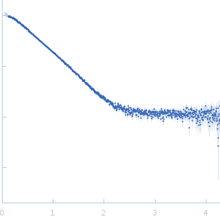
Functional binding region (120-395) of the pneumococcal serine-rich repeat protein (PsrP BR*(120-395))
Mol. type   Protein
Organism   Streptococcus pneumoniae
Olig. state   Monomer
Mon. MW   30.3 kDa
 
UniProt   A0A0H2URK1 (120-395)
Sequence   FASTA