KSHV but not MHV-68 LANA induces a strong bend upon binding to terminal repeat viral DNA.

Ponnusamy R, Petoukhov MV, Correia B, Custodio TF, Juillard F, Tan M, Pires de Miranda M, Carrondo MA, Simas JP, Kaye KM, Svergun DI, McVey CE, Nucleic Acids Res 43(20):10039-54 (2015) Europe PMC

SASDAM8 – MHV-68 LANA

Latency-associated nuclear antigen
MWexperimental 100 kDa
MWexpected 87 kDa
VPorod 117 nm3
log I(s) 4.84×10³ 4.84×10² 4.84×101 4.84×100
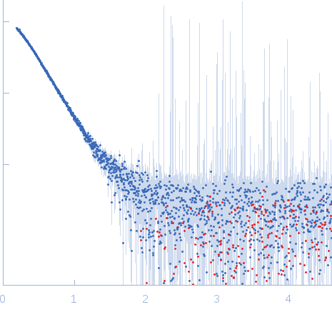
Latency-associated nuclear antigen small angle scattering data  s, nm-1
ln I(s)
Latency-associated nuclear antigen Guinier plot ln 4.85×10³ Rg: 4.2 nm 0 (4.2 nm)-2 s2
(sRg)2I(s)/I(0)
Latency-associated nuclear antigen Kratky plot 1.104 0 3 sRg
p(r)
Latency-associated nuclear antigen pair distance distribution function Rg: 4.4 nm 0 Dmax: 16 nm

Data validation


Fits and models


log I(s)
 s, nm-1
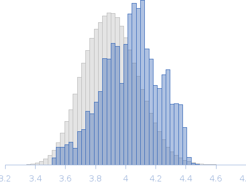
MHV-68 LANA Rg histogram Rg, nm
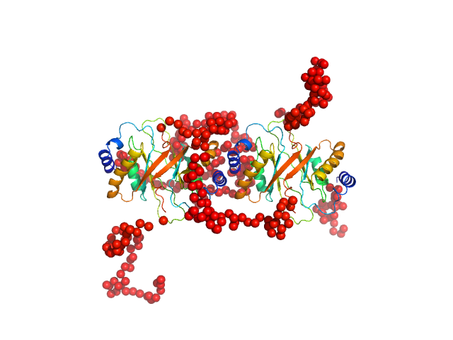
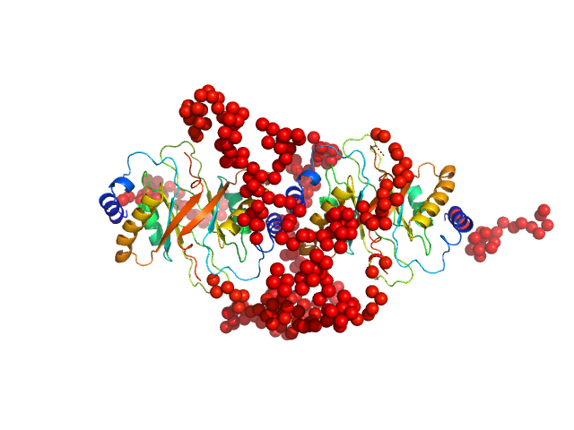
Latency-associated nuclear antigen EOM/RANCH model

Synchrotron SAXS data from solutions of MHV-68 LANA in 25 mM Na/K Phosphate, pH 7.5 were collected on the EMBL P12 beam line at the PETRA III storage ring (DESY; Hamburg, Germany) using a Pilatus 2M detector at a sample-detector distance of 3.1 m and at a wavelength of λ = 0.12 nm (I(s) vs s, where s = 4πsinθ/λ, and 2θ is the scattering angle). One solute concentration of 5.00 mg/ml was measured at 10°C. 20 successive 0.050 second frames were collected. The data were normalized to the intensity of the transmitted beam and radially averaged; the scattering of the solvent-blank was subtracted.

Study of oligomeric states of mLANA and kLANA and their complexes with DNA

Latency-associated nuclear antigen (mLANA)
Mol. type   Protein
Organism   Murid herpesvirus 4
Olig. state   Tetramer
Mon. MW   21.7 kDa
 
UniProt   O41974 (124-316)
Sequence   FASTA