|
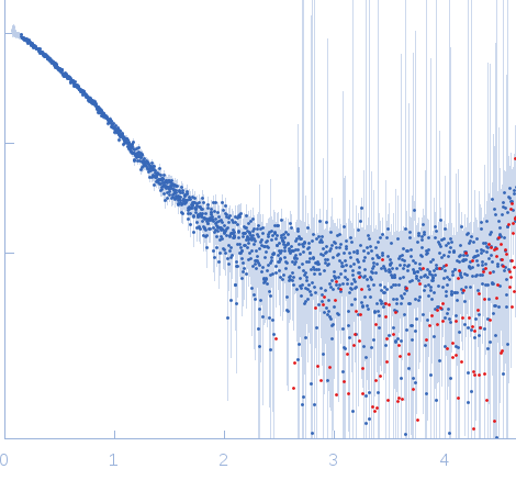
Synchrotron SAXS
data from solutions of
Chitinase 60 from Moritella marina
in
20 mM Tris 200 mM NaCl, pH 8
were collected
on the
EMBL P12 beam line
at the PETRA III storage ring
(DESY; Hamburg, Germany)
using a Pilatus 2M detector
at a sample-detector distance of 3.1 m and
at a wavelength of λ = 0.12 nm
(I(s) vs s, where s = 4πsinθ/λ, and 2θ is the scattering angle).
Solute concentrations ranging between 0.6 and 8.8 mg/ml were measured
at 10°C.
The data were normalized to the intensity of the transmitted beam and radially averaged; the scattering of the solvent-blank was subtracted.
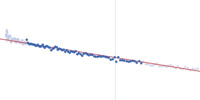
The fit is obtained by the average of three EOM models
|
|
Chitinase 60
|
| Mol. type |
|
Protein |
| Organism |
|
Moritella marina |
| Olig. state |
|
Monomer |
| Mon. MW |
|
60.8 kDa |
| |
| UniProt |
|
B1VBB0
|
| Sequence |
|
FASTA |
| |
|
 s, nm-1
 Rg, nm