X-Ray Solution Scattering Study of Four Escherichia coli Enzymes Involved in Stationary-Phase Metabolism.

Dadinova LA, Shtykova EV, Konarev PV, Rodina EV, Snalina NE, Vorobyeva NN, Kurilova SA, Nazarova TI, Jeffries CM, Svergun DI, PLoS One 11(5):e0156105 (2016) Europe PMC

SASDB33 – Glutamate decarboxylase alpha (GadA) from E. coli

Glutamate decarboxylase alpha (GadA) from E. coli
MWI(0) 249 kDa
MWexpected 53 kDa
VPorod 410 nm3
log I(s) 1.41×10⁴ 1.41×10³ 1.41×10² 1.41×101
Glutamate decarboxylase alpha (GadA) from E. coli small angle scattering data  s, nm-1
ln I(s)
Glutamate decarboxylase alpha (GadA) from E. coli Guinier plot ln 1.41×10⁴ Rg: 4.8 nm 0 (4.8 nm)-2 s2
(sRg)2I(s)/I(0)
Glutamate decarboxylase alpha (GadA) from E. coli Kratky plot 1.104 0 3 sRg

Data validation


Fits and models


log I(s)
 s, nm-1
Glutamate decarboxylase alpha (GadA) from E. coli SASREF MX model

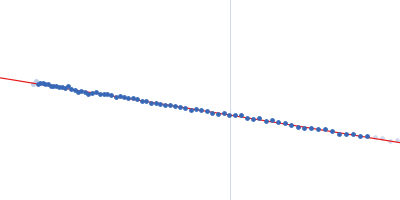
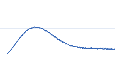
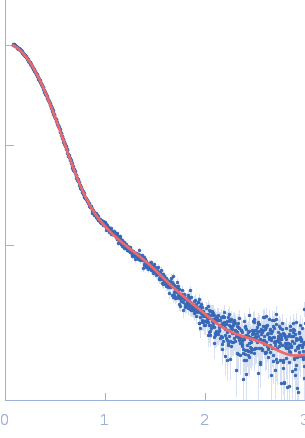
Synchrotron SAXS data from solutions of Glutamate decarboxylase alpha (GadA) from E. coli in 50 mM Tris, pH 7.5 were collected on the P12 beam line of Petra-III (Hamburg, Germany) using a Pilatus 2M detector (I(s) vs s; s = 4π sin θ/λ, where 2θ is the scattering angle and λ=0.124 nm). Different solute concentrations in the range 1.8-8.5 mg/ml were measured using an exposure time of 1 s (recorded as 20 x 0.050 s frames). The data were normalized to the intensity of the transmitted beam and radially averaged and the scattering from the matched solvent-blank was subtracted. The data presented here are from a single concentration scattering curve (8.5 mg/ml).

Under the experimental conditions described above, GadA exists as a mixture in solution of likely hexamers (volume fraction approximately 60%) and disassociated dimers (40%). The models displayed for this entry and associated fit are derived from SASREFMX modelling in P32 symmetry. The final fit to the SAXS data of the mixture was determined using OLIGOMER. The specific volume fraction estimates of the hexamer and three dimers are included in the full entry zip archive.
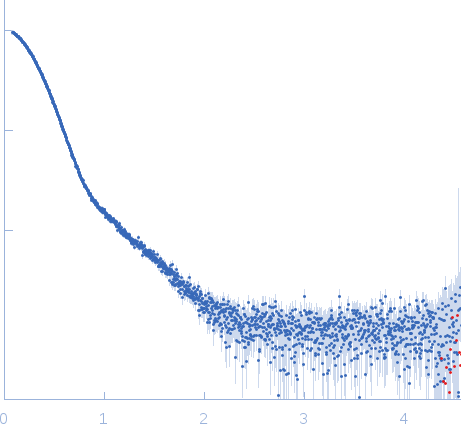
Glutamate decarboxylase alpha (GadA) from E. coli (GadA)
Mol. type   Protein
Organism   Escherichia coli
Olig. state   Monomer
Mon. MW   52.7 kDa
 
UniProt   P69908
Sequence   FASTA
 
PDB ID   1XEY