X-Ray Solution Scattering Study of Four Escherichia coli Enzymes Involved in Stationary-Phase Metabolism.

Dadinova LA, Shtykova EV, Konarev PV, Rodina EV, Snalina NE, Vorobyeva NN, Kurilova SA, Nazarova TI, Jeffries CM, Svergun DI, PLoS One 11(5):e0156105 (2016) Europe PMC

SASDBY2 – Inorganic pyrophosphatase (PPase) from E. coli

Inorganic pyrophosphatase (PPase) from E. coli
MWI(0) 130 kDa
MWexpected 117 kDa
VPorod 166 nm3
log I(s) 6.79×10³ 6.79×10² 6.79×101 6.79×100
Inorganic pyrophosphatase (PPase) from E. coli small angle scattering data  s, nm-1
ln I(s)
Inorganic pyrophosphatase (PPase) from E. coli Guinier plot ln 6.80×10³ Rg: 3.0 nm 0 (3.0 nm)-2 s2
(sRg)2I(s)/I(0)
Inorganic pyrophosphatase (PPase) from E. coli Kratky plot 1.104 0 3 sRg
p(r)
Inorganic pyrophosphatase (PPase) from E. coli pair distance distribution function Rg: 3 nm 0 Dmax: 9 nm

Data validation


Fits and models


log I(s)
 s, nm-1
Inorganic pyrophosphatase (PPase) from E. coli DAMMIN model

log I(s)
 s, nm-1
Inorganic pyrophosphatase (PPase) from E. coli SASREF model

log I(s)
 s, nm-1
Inorganic pyrophosphatase (PPase) from E. coli PDB (PROTEIN DATA BANK) model

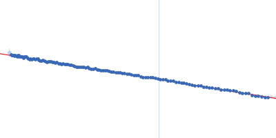
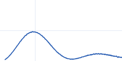
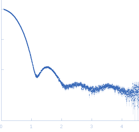
Synchrotron SAXS data from solutions of Inorganic pyrophosphatase (PPase) from E. coli in 50 mM Tris, pH 7.5 were collected on the P12 beam line of Petra-III (Hamburg, Germany) using a Pilatus 2M detector (I(s) vs s; s = 4π sin θ/λ, where 2θ is the scattering angle and λ=0.124 nm). Different solute concentrations in the range 2.8-10.90 mg/ml were measured using an exposure time of 1 s (recorded as 20 x 0.050 s frames). The data were normalized to the intensity of the transmitted beam and radially averaged and the scattering from the matched solvent-blank was subtracted. The data presented here are the scattering intensities derived from the low concentration data set (at 2.8 mg/ml) for s < 0.54 nm-1 merged with the high concentration data for s > 0.54 nm-1 (all data across the concentration series are statistically similar for s > 0.54 nm-1, Correlation Map p = 0.11-1.0).

The models displayed above are, from top to bottom: 1) An individual DAMMIN dummy atom model; 2) SASREF rigid body model with the associated CRYSOL30 fit to the data and; 3) The PPase crystal structure fit to the data (derived from Protein data bank entry 2AUU). All of the individual DAMMIN models, associated fits and averaging can be found in the zip archive for this entry.

Inorganic pyrophosphatase (PPase) from E. coli (PPase)
Mol. type   Protein
Organism   Escherichia coli
Olig. state   Hexamer
Mon. MW   19.6 kDa
 
UniProt   P0A7A9
Sequence   FASTA
 
PDB ID   2AUU