Allosteric communication between DNA-binding and light-responsive domains of diatom class I aureochromes.

Banerjee A, Herman E, Serif M, Maestre-Reyna M, Hepp S, Pokorny R, Kroth PG, Essen LO, Kottke T, Nucleic Acids Res 44(12):5957-70 (2016) Europe PMC

SASDBY3 – Aureochrome 1a bZIP-LOV module: PtAUREO1a bZIP-LOV (Light oxygen voltage) module (light state-TBE)

Aureochrome 1a bZIP-LOV module
MWexperimental 58 kDa
MWexpected 57 kDa
VPorod 115 nm3
log I(s) 5.78×101 5.78×100 5.78×10⁻¹ 5.78×10⁻²
Aureochrome 1a bZIP-LOV module small angle scattering data  s, nm-1
ln I(s)
Aureochrome 1a bZIP-LOV module Guinier plot ln 5.79×101 Rg: 3.4 nm 0 (3.4 nm)-2 s2
(sRg)2I(s)/I(0)
Aureochrome 1a bZIP-LOV module Kratky plot 1.104 0 3 sRg
p(r)
Aureochrome 1a bZIP-LOV module pair distance distribution function Rg: 3.5 nm 0 Dmax: 12.6 nm

Data validation


Fits and models


log I(s)
 s, nm-1
Aureochrome 1a bZIP-LOV module DAMFILT model

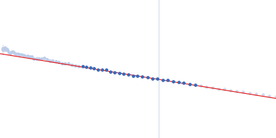
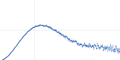
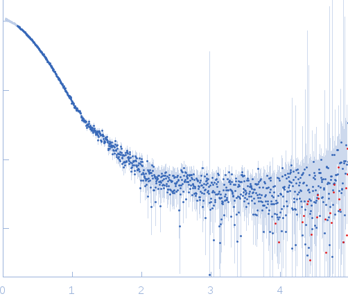
X-ray synchrotron radiation scattering data from solution of PtAUREO1a bZIP-LOV module in 0.5X TBE (50 mM Tris pH 8, 50 mM boric acid, 1 mM EDTA) buffer was collected on the BM29 camera on the storage ring ESRF (Grenoble, France) using a 2D Photon counting Pilatus 1M-W pixel detector (s = 4π sin θ/λ, where 2θ is the scattering angle). Ten successive 20 second frames were collected. The data were normalized to the intensity of the transmitted beam and radially averaged; the scattering of the solvent-blank was subtracted. Solution scattering data of bZIP-LOV module in the light state was performed at 1.25mg/mL concentration at 4 degrees centigrade. The modelling analysis was performed using the ATSAS package. The displayed model is an averaged and volume corrected dummy atom representation (DAMFILT). For further sequence information refer to the Joint Genome Institute (JGI) entry JGI-49116.

Aureochrome 1a bZIP-LOV module (PtAUREO1a bZIP-LOV)
Mol. type   Protein
Organism   Phaeodactylum tricornutum
Olig. state   Dimer
Mon. MW   28.7 kDa
Sequence   FASTA