The Two-State Prehensile Tail of the Antibacterial Toxin Colicin N.

Johnson CL, Solovyova AS, Hecht O, Macdonald C, Waller H, Grossmann JG, Moore GR, Lakey JH, Biophys J 113(8):1673-1684 (2017) Europe PMC

SASDC53 – Colicin N Translocation domain

Colicin N Translocation domain
MWI(0) 19 kDa
MWexpected 10 kDa
VPorod 22 nm3
log I(s) 2.29×101 2.29×100 2.29×10⁻¹ 2.29×10⁻²
Colicin N Translocation domain small angle scattering data  s, nm-1
ln I(s)
Colicin N Translocation domain Guinier plot ln 2.30×101 Rg: 2.8 nm 0 (2.8 nm)-2 s2
(sRg)2I(s)/I(0)
Colicin N Translocation domain Kratky plot 1.104 0 3 sRg
p(r)
Colicin N Translocation domain pair distance distribution function Rg: 2.9 nm 0 Dmax: 11.4 nm

Data validation


Fits and models


log I(s)
 s, nm-1
Colicin N Translocation domain Rg histogram Rg, nm
Colicin N Translocation domain EOM/GAJOE model

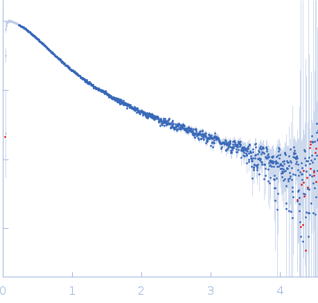
Synchrotron SAXS data from solutions of Colicin N Translocation domain in 50 mM Na-Phosphate 300 mM NaCl, pH 7.6 were collected on the BM29 beam line at the ESRF (Grenoble, France) using a Pilatus 1M detector at a sample-detector distance of 2.9 m and at a wavelength of λ = 0.15 nm (I(s) vs s, where s = 4πsinθ/λ and 2θ is the scattering angle). Solute concentrations ranging between 0.4 and 6.3 mg/ml were measured at 4°C. 10 successive 2 second frames were collected. The data were normalized to the intensity of the transmitted beam and radially averaged; the scattering of the solvent-blank was subtracted and the different curves were scaled for protein concentration.

The results obtained from the bayesapp estimation of distribution functions from small-angle scattering data (http://www.bayesapp.org/) are included in the full entry zip archive.

Tags: idp
Colicin N Translocation domain (ColN-T)
Mol. type   Protein
Organism   Escherichia coli
Olig. state   Monomer
Mon. MW   10.0 kDa
 
UniProt   P08083 (1-90)
Sequence   FASTA