Revealing the distinct folding phases of an RNA three-helix junction.

Plumridge A, Katz AM, Calvey GD, Elber R, Kirmizialtin S, Pollack L, Nucleic Acids Res 46(14):7354-7365 (2018) Europe PMC

SASDCD4 – Truncated P5abc subdomain from tetrahymena ribozyme: Time-resolved 1000ms

Truncated P5abc subdomain from tetrahymena ribozyme
MWexperimental 24 kDa
MWexpected 18 kDa
log I(s) 9.40×10⁻¹ 9.40×10⁻² 9.40×10⁻³ 9.40×10⁻⁴
Truncated P5abc subdomain from tetrahymena ribozyme small angle scattering data  s, nm-1
ln I(s)
Truncated P5abc subdomain from tetrahymena ribozyme Guinier plot ln 9.40×10⁻¹ Rg: 2.2 nm 0 (2.2 nm)-2 s2
(sRg)2I(s)/I(0)
Truncated P5abc subdomain from tetrahymena ribozyme Kratky plot 1.104 0 3 sRg
p(r)
Truncated P5abc subdomain from tetrahymena ribozyme pair distance distribution function Rg: 2.2 nm 0 Dmax: 6.8 nm

Data validation


There are no models related to this curve.

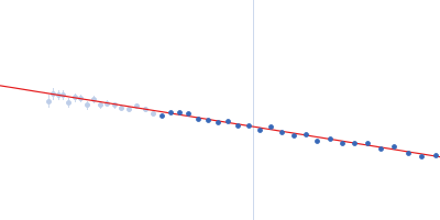
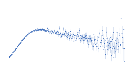
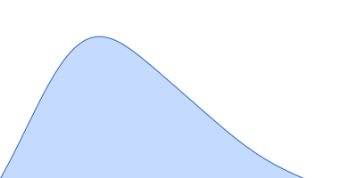
Synchrotron SAXS data from solutions of Truncated P5abc subdomain from tetrahymena ribozyme: (Time-resolved 1000ms) in 10mM KMOPS 20mM KCl 1mM MgCl2 20uM EDTA, pH 7 were collected on the G1 beam line at the Cornell High Energy Synchrotron Source (CHESS; Ithaca, NY, USA) using a CCD Finger Lakes CCD detector at a sample-detector distance of 1.7 m and at a wavelength of λ = 0.109 nm (I(s) vs s, where s = 4πsinθ/λ and 2θ is the scattering angle). One solute concentration of 1.50 mg/ml was measured at 20°C. 50 successive 5 second frames were collected. The data were normalized to the intensity of the transmitted beam and radially averaged; the scattering of the solvent-blank was subtracted.
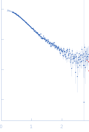
Truncated P5abc subdomain from tetrahymena ribozyme (tp5abc)
Mol. type   RNA
Olig. state   Monomer
Mon. MW   18.2 kDa
Sequence   FASTA