2017 publication guidelines for structural modelling of small-angle scattering data from biomolecules in solution: an update.

Trewhella J, Duff AP, Durand D, Gabel F, Guss JM, Hendrickson WA, Hura GL, Jacques DA, Kirby NM, Kwan AH, Pérez J, Pollack L, Ryan TM, Sali A, Schneidman-Duhovny D, Schwede T, Svergun DI, Sugiyama M, Tainer JA, Vachette P, Westbrook J, Whitten AE, Acta Crystallogr D Struct Biol 73(Pt 9):710-728 (2017) Europe PMC

SASDCK2 – Glucose Isomerase - Streptomyces rubiginosus

Xylose isomerase
MWI(0) 183 kDa
MWexpected 173 kDa
VPorod 229 nm3
log I(s) 3.70×10⁻² 3.70×10⁻³ 3.70×10⁻⁴ 3.70×10⁻⁵
Xylose isomerase small angle scattering data  s, nm-1
ln I(s)
Xylose isomerase Guinier plot ln 3.70×10⁻² Rg: 3.3 nm 0 (3.3 nm)-2 s2
(sRg)2I(s)/I(0)
Xylose isomerase Kratky plot 1.104 0 3 sRg
p(r)
Xylose isomerase pair distance distribution function Rg: 3.3 nm 0 Dmax: 9.2 nm

Data validation


Fits and models


log I(s)
 s, nm-1
Xylose isomerase DAMMIN model

log I(s)
 s, nm-1
Xylose isomerase NONE model

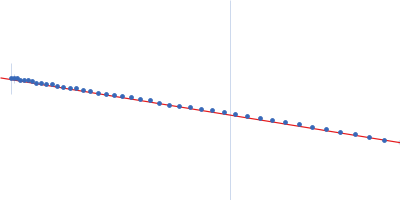
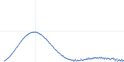
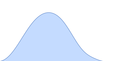
Synchrotron SAXS data from solutions of glucoes isomerase in 25 mM MOPS, 250 mM NaCl, 50 mM KCl, 2 mM TCEP, 0.1% NaN3, pH 7.5 were collected on the SAXS/WAXS beam line at Australian Synchrotron (Melbourne, Australia) using a Pilatus 1M detector at a sample-detector distance of 2.7 m and at a wavelength of λ = 0.10332 nm (I(s) vs s, where s = 4πsinθ/λ and 2θ is the scattering angle). Scattering data are on an absolute scale (cm-1) set by reference to the scattering from pure H2O. I(0) values must be corrected for the shear flow sample cell by a 2.05 multiplicative factor. Data were acquired in SEC-SAXS mode, summed from 21 measurement frames where Rg was flat and at a maximum. The average concentration over the frames was 0.18 mg/mL, spanning values from 0.06 - 0.34 mg/mL. Sample concentrations were measured by A280 (extinction coefficient for 1 mg/mL, 10 mm pathlength; E0.1% = 1.079), corrected for the 3.1 mm path-length of the UV measurement cell. MULCh was used to calculate the partial specific volume (0.732 mL/g) and X-ray contrast (Δρ = 2.833e10 cm-2). The protein is a tetramer in the solution conditions measured.

Storage temperature = UNKNOWN
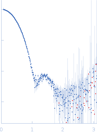
Tags: benchmark
Xylose isomerase (Glucose Isomerase)
Mol. type   Protein
Organism   Streptomyces rubiginosus
Olig. state   Tetramer
Mon. MW   43.2 kDa
 
UniProt   P24300 (1-388)
Sequence   FASTA
 
PDB ID   1OAD