The structure of SALM5 suggests a dimeric assembly for the presynaptic RPTP ligand recognition.

Karki S, Paudel P, Sele C, Shkumatov AV, Kajander T, Protein Eng Des Sel (2018) Europe PMC

SASDD23 – Leucine-rich repeat and fibronectin type-III domain-containing protein 5: Fragment SALM5 LRR-Ig

Leucine-rich repeat and fibronectin type-III domain-containing protein 5
MWexperimental 101 kDa
MWexpected 82 kDa
VPorod 155 nm3
log I(s) 6.72×10⁻³ 6.72×10⁻⁴ 6.72×10⁻⁵ 6.72×10⁻⁶
Leucine-rich repeat and fibronectin type-III domain-containing protein 5 small angle scattering data  s, nm-1
ln I(s)
Leucine-rich repeat and fibronectin type-III domain-containing protein 5 Guinier plot ln 6.72×10⁻³ Rg: 3.6 nm 0 (3.6 nm)-2 s2
(sRg)2I(s)/I(0)
Leucine-rich repeat and fibronectin type-III domain-containing protein 5 Kratky plot 1.104 0 3 sRg
p(r)
Leucine-rich repeat and fibronectin type-III domain-containing protein 5 pair distance distribution function Rg: 3.7 nm 0 Dmax: 13.5 nm

Data validation


Fits and models


log I(s)
 s, nm-1
Leucine-rich repeat and fibronectin type-III domain-containing protein 5: Fragment SALM5 LRR-Ig Rg histogram Rg, nm
Leucine-rich repeat and fibronectin type-III domain-containing protein 5 EOM/RANCH model

log I(s)
 s, nm-1
Leucine-rich repeat and fibronectin type-III domain-containing protein 5 DAMMIN model

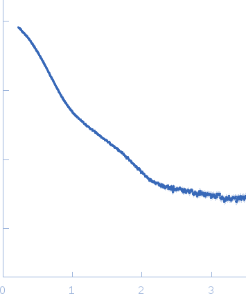
Synchrotron SAXS data from solutions of Leucine-rich repeat and fibronectin type-III domain-containing protein 5 (fragment SALM5 (LRR-Ig)) in 30 mM Tris-Cl, 150 mM NaCl, 3% v/v glycerol, pH 7.5, were collected on the B21 beam line at the Diamond Light Source storage ring (Oxfordshire, UK) using a Pilatus 2M detector at a sample-detector distance of 4.0 m and at a wavelength of λ = 0.1005 nm (l(s) vs s, where s = 4πsinθ/λ, and 2θ is the scattering angle). Solute concentrations ranging between 1 and 5 mg/ml were measured at 15°C. 18 successive 10 second frames were collected. The data were normalized to the intensity of the transmitted beam and radially averaged; the scattering of the solvent-blank was subtracted. The low angle data collected at lower concentration were merged with the highest concentration high angle data to yield the final composite scattering curve.

Leucine-rich repeat and fibronectin type-III domain-containing protein 5 (LRR-Ig-FN)
Mol. type   Protein
Organism   Mus musculus
Olig. state   Dimer
Mon. MW   41.0 kDa
 
UniProt   Q8BXA0 (18-376)
Sequence   FASTA