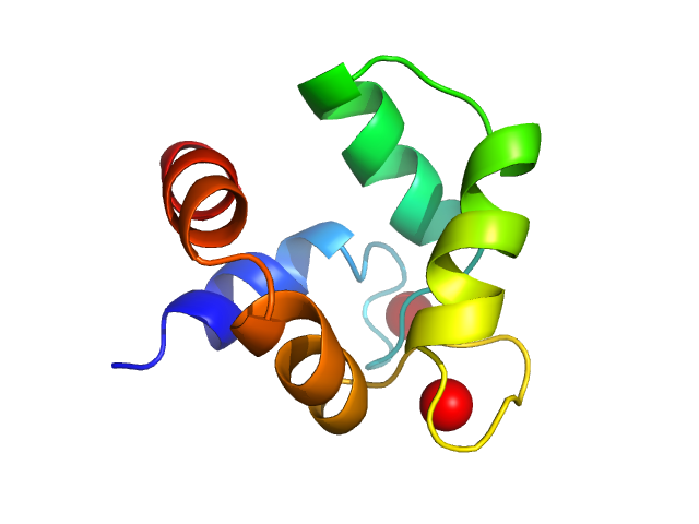
Solution structures of polcalcin Phl p 7 in three ligation states: Apo-, hemi-Mg2+-bound, and fully Ca2+-bound.

Henzl MT, Sirianni AG, Wycoff WG, Tan A, Tanner JJ, Proteins 81(2):300-15 (2013) Europe PMC

SASDDJ2 – Calcium-bound polcalcin Phl p 7

polcalcin Phl p 7
MWexperimental 9 kDa
MWexpected 9 kDa
VPorod 14 nm3
log I(s) 5.25×101 5.25×100 5.25×10⁻¹ 5.25×10⁻²
polcalcin Phl p 7 small angle scattering data  s, nm-1
ln I(s)
polcalcin Phl p 7 Guinier plot ln 5.25×101 Rg: 1.3 nm 0 (1.3 nm)-2 s2
(sRg)2I(s)/I(0)
polcalcin Phl p 7 Kratky plot 1.104 0 3 sRg
p(r)
polcalcin Phl p 7 pair distance distribution function Rg: 1.3 nm 0 Dmax: 3.6 nm

Data validation


Fits and models


log I(s)
 s, nm-1
polcalcin Phl p 7 MES-FOXS model

Synchrotron SAXS data from solutions of calcium-bound polcalcin Phl p 7 in 0.15 M NaCl, 0.025 M HEPES, 100 uM Ca2+, pH 7.4, were collected on the 12.3.1 SIBYLS beam line at the Advanced Light Source storage ring (ALS; Berkeley, CA, USA) using a MAR 165 CCD detector at a wavelength of λ = 0.1127 nm (l(s) vs s, where s = 4πsinθ/λ, and 2θ is the scattering angle). Solute concentrations ranging between 5 and 15 mg/ml were measured at 10°C. The data were normalized to the intensity of the transmitted beam and radially averaged; the scattering of the solvent-blank was subtracted.

Storage temperature = UNKNOWN. Sample detector distance = UNKNOWN. X-ray Exposure time = UNKNOWN. Number of frames = UNKNOWN

polcalcin Phl p 7
Mol. type   Protein
Organism   Phleum pratense
Olig. state   Monomer
Mon. MW   8.5 kDa
 
UniProt   O82040
Sequence   FASTA