Inhibitor-induced dimerization of an essential oxidoreductase from African Trypanosomes.

Wagner A, Le TA, Brennich M, Klein P, Bader N, Diehl E, Paszek D, Weickhmann AK, Dirdjaja N, Krauth-Siegel RL, Engels B, Opatz T, Schindelin H, Hellmich UA, Angew Chem Int Ed Engl (2019) Europe PMC

SASDE94 – Tryparedoxin I109A, reduced state

Tryparedoxin I109A
MWexperimental 15 kDa
MWexpected 16 kDa
VPorod 25 nm3
log I(s) 2.62×100 2.62×10⁻¹ 2.62×10⁻² 2.62×10⁻³
Tryparedoxin I109A small angle scattering data  s, nm-1
ln I(s)
Tryparedoxin I109A Guinier plot ln 2.62×100 Rg: 1.6 nm 0 (1.6 nm)-2 s2
(sRg)2I(s)/I(0)
Tryparedoxin I109A Kratky plot 1.104 0 3 sRg
p(r)
Tryparedoxin I109A pair distance distribution function Rg: 1.6 nm 0 Dmax: 6 nm

Data validation


Fits and models


log I(s)
 s, nm-1
Tryparedoxin I109A PDB (PROTEIN DATA BANK) model
Tryparedoxin I109A OTHER model

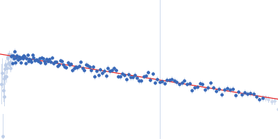
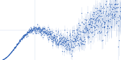
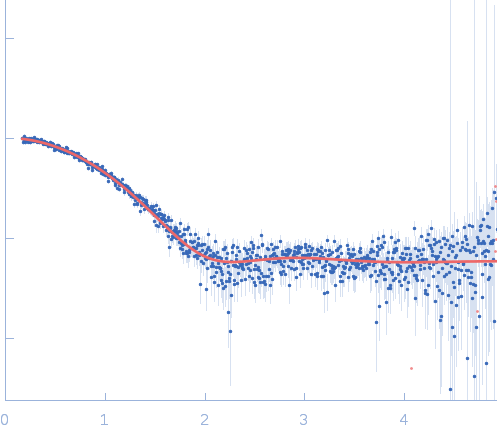
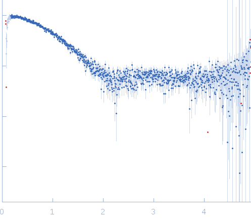
Synchrotron SAXS data from solutions of Tryparedoxin I109A, reduced state in 10 mM HEPES pH 7.5, 50 mM NaCl, pH 7.5 were collected on the BM29 beam line at the ESRF (Grenoble, France) using a Dectris Pilatus 1M detector at a sample-detector distance of 2.9 m and at a wavelength of λ = 0.099 nm (l(s) vs s, where s = 4πsinθ/λ, and 2θ is the scattering angle). The data were collected using in-line size exclusion chromatography (SEC)-SAXS at 20°C.

SEC column type: GE Healthcare S75 3.2/300; Sample Injection Concentration = 10 mg/mL; Column flow-rate = 0.1 mL/min.

Tryparedoxin I109A
Mol. type   Protein
Organism   Trypanosoma brucei brucei
Olig. state   Monomer
Mon. MW   15.8 kDa
 
UniProt   O77404
Sequence   FASTA
 
PDB ID   6GXY