Marsin S,
Adam Y,
Cargemel C,
Andreani J,
Baconnais S,
Legrand P,
Li de la Sierra-Gallay I,
Humbert A,
Aumont-Nicaise M,
Velours C,
Ochsenbein F,
Durand D,
Le Cam E,
Walbott H,
Possoz C,
Quevillon-Cheruel S,
Ferat JL,
Nucleic Acids Res
(2021)
Europe PMC
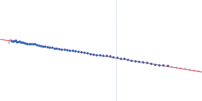
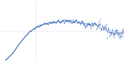
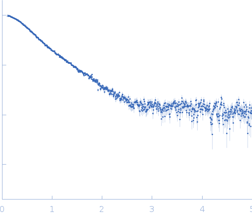
Synchrotron SAXS data from solutions of DciA in 20 mM Tris-HCl, 100 mM NaCl, pH 7.5 were collected on the SWING beam line at SOLEIL (Saint-Aubin, France) using a AVIEX PCCD170170 detector at a sample-detector distance of 1.8 m and at a wavelength of λ = 0.1033 nm (I(s) vs s, where s = 4πsinθ/λ, and 2θ is the scattering angle). In-line size-exclusion chromatography (SEC) SAS was employed. The SEC parameters were as follows: A 95.00 μl sample at 5.7 mg/ml was injected at a 0.50 ml/min flow rate onto a GE Superose 6 10/300 column at 15°C. 254 successive 2 second frames were collected. The data were normalized to the intensity of the transmitted beam and radially averaged; the scattering of the solvent-blank was subtracted.
The scattered intensities were displayed on an absolute scale (cm-1) using the scattering of water. Frames were examined individually using the US-SOMO HPLC module and 26 identical frames obtained around the maximum of the elution peak were averaged and further processed.
 s, nm-1