|
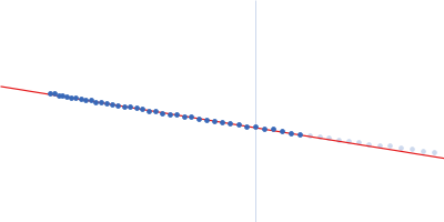
Synchrotron SAXS
data from solutions of
MvaT (low salt data set)
in
20 mM Bis-Tris 50 mM KCl, pH 6
were collected
on the
BM29 beam line
at the ESRF storage ring
(Grenoble, France)
using a Pilatus 1M detector
at a sample-detector distance of 2.8 m and
at a wavelength of λ = 0.099 nm
(I(s) vs s, where s = 4πsinθ/λ, and 2θ is the scattering angle).
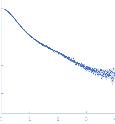
In-line size-exclusion chromatography (SEC) SAS was employed. The SEC parameters were as follows: A 250.00 μl sample
at 11 mg/ml was injected
onto a GE Superdex 75 Increase 10/300 column
at 20°C.
The data were normalized to the intensity of the transmitted beam and radially averaged; the scattering of the solvent-blank was subtracted.
The protein employed in the SAS experiment contains three point mutations: K31C, F36D, and M44D. K31C was introduced to introduce a paramagnetic label, while F36D and M44D were introduced to abolish higher-order oligomerisation of the protein.
|
|
 s, nm-1
![MvaT(mutant) OTHER [STATIC IMAGE] model](/media//pdb_file/SASDGS5_fit1_model1.png "Static model image")