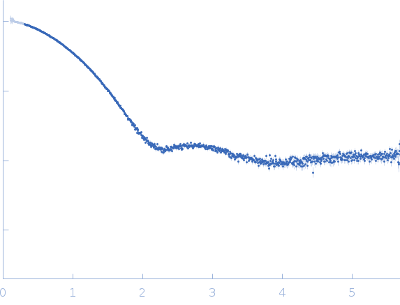
Structural and Biophysical Analysis of the CLCA1 VWA Domain Suggests Mode of TMEM16A Engagement.

Berry KN, Brett TJ, Cell Rep 30(4):1141-1151.e3 (2020) Europe PMC

SASDH24 – Calcium-Activated Chloride Channel Regulator 1 VWA domain

Calcium-activated chloride channel regulator 1
MWexperimental 16 kDa
MWexpected 20 kDa
log I(s) 1.45×101 1.45×100 1.45×10⁻¹ 1.45×10⁻²
Calcium-activated chloride channel regulator 1 small angle scattering data  s, nm-1
ln I(s)
Calcium-activated chloride channel regulator 1 Guinier plot ln 1.46×101 Rg: 1.8 nm 0 (1.8 nm)-2 s2
(sRg)2I(s)/I(0)
Calcium-activated chloride channel regulator 1 Kratky plot 1.104 0 3 sRg
p(r)
Calcium-activated chloride channel regulator 1 pair distance distribution function Rg: 1.8 nm 0 Dmax: 6.6 nm

Data validation


Fits and models


log I(s)
 s, nm-1
Calcium-activated chloride channel regulator 1 DAMFILT model

Synchrotron SAXS data from solutions of Calcium-Activated Chloride Channel Regulator 1 VWA domain in 20 mM HEPES, 150 mM NaCl, 2% glycerol, pH 7.4 were collected on the 12-ID-B SAXS/WAXS beam line at the Advanced Photon Source (APS), Argonne National Laboratory storage ring (Lemont, IL, USA) using a Pilatus 2M detector at a sample-detector distance of 1.5 m and at a wavelength of λ = 0.127 nm (I(s) vs s, where s = 4πsinθ/λ, and 2θ is the scattering angle). Solute concentrations ranging between 2.0 and 8.2 mg/ml were measured at 10°C. 32 successive 0.300 second frames were collected. The data were normalized to the intensity of the transmitted beam and radially averaged; the scattering of the solvent-blank was subtracted. The low angle data collected at lower concentration were merged with the highest concentration high angle data to yield the final composite scattering curve.

Calcium-activated chloride channel regulator 1 (CLCA1)
Mol. type   Protein
Organism   Homo sapiens
Olig. state   Monomer
Mon. MW   19.8 kDa
 
UniProt   A8K7I4 (302-476)
Sequence   FASTA