A key interaction with RPA orients XPA in NER complexes.

Topolska-Woś AM, Sugitani N, Cordoba JJ, Le Meur KV, Le Meur RA, Kim HS, Yeo JE, Rosenberg D, Hammel M, Schärer OD, Chazin WJ, Nucleic Acids Res (2020) Europe PMC

SASDH54 – 5' Complex of XPA-DBD with RPA70AB

DNA repair protein complementing XP-A cells
Replication protein A 70 kDa DNA-binding subunit
5-prime Nucleotide Excision Repair Junction Model Substrate
MWI(0) 58 kDa
MWexpected 55 kDa
VPorod 87 nm3
log I(s) 1.66×101 1.66×100 1.66×10⁻¹ 1.66×10⁻²
DNA repair protein complementing XP-A cells Replication protein A 70 kDa DNA-binding subunit 5-prime Nucleotide Excision Repair Junction Model Substrate small angle scattering data  s, nm-1
ln I(s)
DNA repair protein complementing XP-A cells Replication protein A 70 kDa DNA-binding subunit 5-prime Nucleotide Excision Repair Junction Model Substrate Guinier plot ln 1.67×101 Rg: 2.9 nm 0 (2.9 nm)-2 s2
(sRg)2I(s)/I(0)
DNA repair protein complementing XP-A cells Replication protein A 70 kDa DNA-binding subunit 5-prime Nucleotide Excision Repair Junction Model Substrate Kratky plot 1.104 0 3 sRg
p(r)
DNA repair protein complementing XP-A cells Replication protein A 70 kDa DNA-binding subunit 5-prime Nucleotide Excision Repair Junction Model Substrate pair distance distribution function Rg: 3.1 nm 0 Dmax: 97 nm

Data validation


Fits and models


log I(s)
 s, nm-1
DNA repair protein complementing XP-A cells Replication protein A 70 kDa DNA-binding subunit 5-prime Nucleotide Excision Repair Junction Model Substrate HADDOCK model

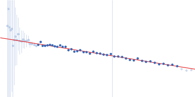
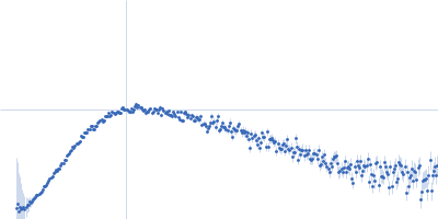
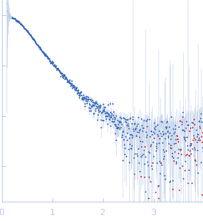
Synchrotron SAXS data from solutions of 5' Complex of XPA-DBD with RPA70AB in 20 mM Tris, 150 mM NaCl, 2% glycerol, 1 mM DTT, pH 7.5 were collected on the 12.3.1 (SIBYLS) beam line at the Advanced Light Source (ALS; Berkeley, CA, USA) using a Pilatus3 X 2M detector at a sample-detector distance of 1.5 m and at a wavelength of λ = 0.103 nm (I(s) vs s, where s = 4πsinθ/λ, and 2θ is the scattering angle). In-line size-exclusion chromatography (SEC) SAS was employed. The SEC parameters were as follows: A 50.00 μl sample was injected at a 0.50 ml/min flow rate onto a Shodex LW-803 column . Data measured from solute-free fractions and the sample-elution peak were normalized to the intensity of the transmitted beam and radially averaged. The scattering of the solvent-blank was subtracted from the sample peak data produce the data displayed in this entry.

Cell temperature = UNKNOWN. Storage temperature = UNKNOWN. Number of frames = UNKNOWN

DNA repair protein complementing XP-A cells
Mol. type   Protein
Organism   Homo sapiens
Olig. state   Monomer
Mon. MW   17.2 kDa
 
UniProt   P23025 (98-239)
Sequence   FASTA
 
Replication protein A 70 kDa DNA-binding subunit (RPA70AB)
Mol. type   Protein
Organism   Homo sapiens
Olig. state   Monomer
Mon. MW   26.7 kDa
 
UniProt   P27694 (183-420)
Sequence   FASTA
 
5-prime Nucleotide Excision Repair Junction Model Substrate (5-prime jxn)
Mol. type   DNA
Olig. state   Monomer
Mon. MW   10.9 kDa
Sequence   FASTA