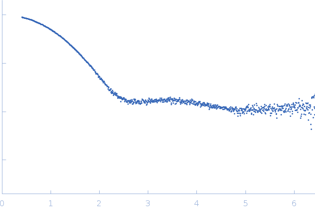
Synchrotron SAXS data from a solution of 7.00 mg/ml human Hen Egg White Lysozyme (HEWL) in 40 mM sodium acetate pH 3.8, 150 mM NaCl, 100 mM 5-methyl uridine was collected on the BL11-NCD-SWEET beam line at ALBA Synchrotron using a Pilatus 1M photon-counting detector. The beam at sample position was 0.35 mm (horizontal) × 0.38 mm (vertical), the incident photon flux was 7.6×10e11 photons/s at 12.4 keV (λ =0.0999 nm) and the sample to detector distance was 2.291 m. 2000 successive 0.1 second frames were collected and data were normalized to the intensity of the transmitted beam and radially averaged. The current buffer-subtracted scattering profile corresponds to the average of 225 collected frames. The absorbed dose by the sample was 13.50 kGy.
Sample temperature: 25°C.
CAUTION! The scattering intensities do not have associated errors.
 s, nm-1