Tandem RNA binding sites induce self-association of the stress granule marker protein TIA-1.

Loughlin FE, West DL, Gunzburg MJ, Waris S, Crawford SA, Wilce MCJ, Wilce JA, Nucleic Acids Res (2021) Europe PMC

SASDJL5 – Nucleolysin TIA-1 isoform p40 (TIA-1) bound to UC1 RNA

Nucleolysin TIA-1 isoform p40
UC1
MWexperimental 42 kDa
MWexpected 24 kDa
VPorod 77 nm3
log I(s) 6.90×10⁻² 6.90×10⁻³ 6.90×10⁻⁴ 6.90×10⁻⁵
Nucleolysin TIA-1 isoform p40 UC1 small angle scattering data  s, nm-1
ln I(s)
Nucleolysin TIA-1 isoform p40 UC1 Guinier plot ln 6.90×10⁻² Rg: 3.3 nm 0 (3.3 nm)-2 s2
(sRg)2I(s)/I(0)
Nucleolysin TIA-1 isoform p40 UC1 Kratky plot 1.104 0 3 sRg
p(r)
Nucleolysin TIA-1 isoform p40 UC1 pair distance distribution function Rg: 3.5 nm 0 Dmax: 14.4 nm

Data validation


There are no models related to this curve.

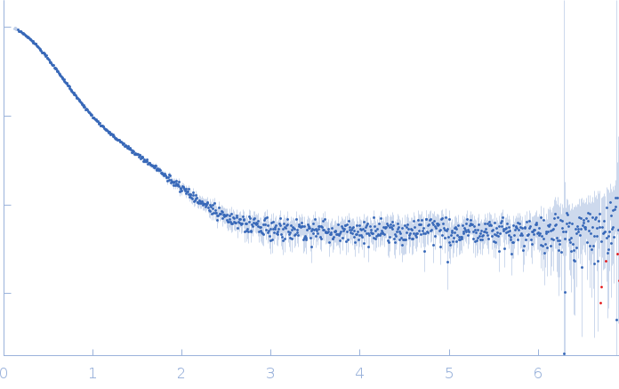
Synchrotron SAXS data from solutions of TIA-1 bound to UC1 RNA in 20 mM HEPES, 100 mM NaCl, 3% v/v glycerol, pH 7 were collected on the SAXS/WAXS beam line at the Australian Synchrotron (Melbourne, Australia) using a Pilatus3 S 2M detector at a sample-detector distance of 1.4 m and at a wavelength of λ = 0.10332 nm (I(s) vs s, where s = 4πsinθ/λ, and 2θ is the scattering angle). 30 successive 1 second frames were collected from a samples at 15°C. The data were normalized to the intensity of the transmitted beam and radially averaged; the scattering of the solvent-blank was subtracted.

Concentration = UNKNOWN

Nucleolysin TIA-1 isoform p40 (TIA-1)
Mol. type   Protein
Organism   Homo sapiens
Olig. state   Monomer
Mon. MW   21.3 kDa
 
UniProt   P31483-2 (93-274)
Sequence   FASTA
 
UC1 (UC1)
Mol. type   RNA
Organism   synthetic construct
Olig. state   Monomer
Mon. MW   3.1 kDa
Sequence   FASTA