Dmax unknown – experimental data range validation not possible.
There are no models related to this curve.
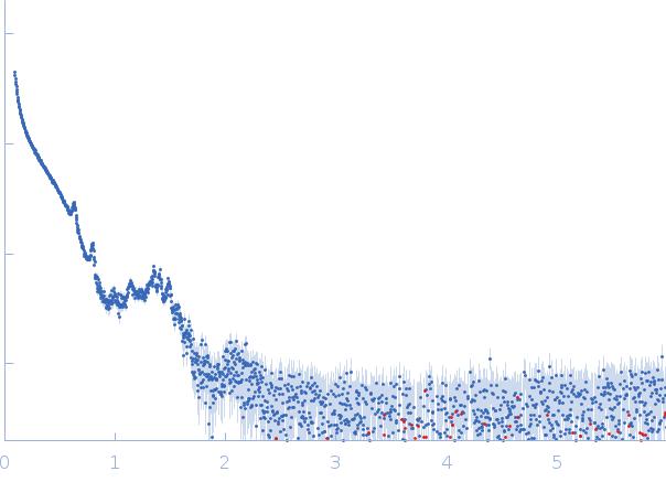
Synchrotron SAXS data from Dps-DNA co-crystalls, in cellulo, were collected on the EMBL-P12 BioSAXS beamline at the PETRAIII storage ring (DESY, Hamburg, Germany) equipped with a robotic sample changer and a 2D photon counting pixel X-ray detector (Pilatus 6M). The scattering intensity I(s) was recorded in the range of the momentum transfer 0.02 < s < 6.0 nm-1, where s = 4πsinθ/λ, and 2θ is the scattering angle, λ= 0.124 nm is the X-ray wavelength. The measurements were carried out in 50 mM Tris-HCl, pH 8, 50 mM NaCl, 0.5 mM EDTA buffer. The data were normalized to the intensity of the transmitted beam and radially averaged; the scattering of the solvent-blank was subtracted.
NOTE: Data validation metrics do not apply for this entry (Rg, I(0), MW, etc). The quoted 'experimental MW' is that of the dodecameric protein calculated from the amino acid sequence. The monomer MW is quoted at 19 kDa.
 s, nm-1