A round-robin approach provides a detailed assessment of biomolecular small-angle scattering data reproducibility and yields consensus curves for benchmarking

Trewhella J, Vachette P, Bierma J, Blanchet C, Brookes E, Chakravarthy S, Chatzimagas L, Cleveland T, Cowieson N, Crossett B, Duff A, Franke D, Gabel F, Gillilan R, Graewert M, Grishaev A, Guss J, Hammel M, Hopkins J, Huang Q, Hub J, Hura G, Irving T, Jeffries C, Jeong C, Kirby N, Krueger S, Martel A, Matsui T, Li N, Pérez J, Porcar L, Prangé T, Rajkovic I, Rocco M, Rosenberg D, Ryan T, Seifert S, Sekiguchi H, Svergun D, Teixeira S, Thureau A, Weiss T, Whitten A, Wood K, Zuo X, Acta Crystallographica Section D Structural Biology 78(11) (2022) DOI

SASDP55 – Consensus SANS Profile - Xylose isomerase in H2O buffer

Xylose isomerase
MWexperimental 172 kDa
MWexpected 173 kDa
log I(s) 6.11×10⁻¹ 6.11×10⁻² 6.11×10⁻³ 6.11×10⁻⁴
Xylose isomerase small angle scattering data  s, nm-1
ln I(s)
Xylose isomerase Guinier plot ln 6.12×10⁻¹ Rg: 3.3 nm 0 (3.3 nm)-2 s2
(sRg)2I(s)/I(0)
Xylose isomerase Kratky plot 1.104 0 3 sRg
p(r)
Xylose isomerase pair distance distribution function Rg: 3.2 nm 0 Dmax: 9.7 nm

Data validation


Fits and models


log I(s)
 s, nm-1
Xylose isomerase PDB (PROTEIN DATA BANK) model

log I(s)
 s, nm-1
Xylose isomerase PDB (PROTEIN DATA BANK) model

log I(s)
 s, nm-1
Xylose isomerase OTHER model

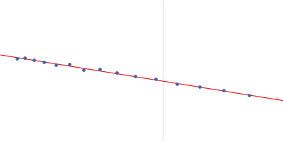
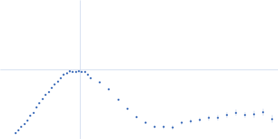
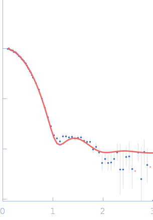
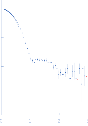
The consensus SANS profile for xylose isomerase in H2O buffer was obtained in a two-step process. First, a consensus batch data set for seven samples with protein concentrations 0.95 – 2.4 mg/mL was generated using the datcombine tool (ATSAS 3.1.0) with both outlier- and error-filters applied. The resulting curve was subsequently merged with data for s > 0.4 nm-1 measured from two samples at 6.8 mg/mL. All data were independent measurements, and no individual measurement was represented more than once in the contributing scattering profile set. The buffer was 50 mM Tris, pH 7.5, 100 mM NaCl. The xylose isomerase atomistic model for CRYSON and Pepsi-SANS fits was the PDB ID 1MNZ tetramer with an N-terminal Met added using PyMol, and small-molecule crystallisation agents removed. Custom WAXSiS-type calculations (with neutron scattering factors and Gromacs software) used the same coordinates and added explicit waters and appropriate number of ions for the MD calculations.

The data input to datcombine are made available for download in the associated zip file, including also the unsubtracted 6 column format data with resolution data. Model fits are shown in order (top to bottom): CRYSON, Pepsi-SANS, and custom WAXSiS (with neutron scattering factors).

Tags: benchmark
Xylose isomerase
Mol. type   Protein
Organism   Streptomyces rubiginosus
Olig. state   Tetramer
Mon. MW   43.2 kDa
 
UniProt   P24300 (1-388)
Sequence   FASTA
 
PDB ID   1MNZ
 
PDB ID   1MNZ