Asymmetric horseshoe-like assembly of peroxisomal yeast oxalyl-CoA synthetase.

Bürgi J, Lill P, Giannopoulou EA, Jeffries CM, Chojnowski G, Raunser S, Gatsogiannis C, Wilmanns M, Biol Chem (2023) Europe PMC

SASDPY3 – Point mutant K352D oxalyl-CoA synthetase Pcs60p (0.5 mg/ml) - dimer

Oxalate--CoA ligase (K352D)
MWexperimental 115 kDa
MWexpected 121 kDa
VPorod 194 nm3
log I(s) 5.20×10⁻² 5.20×10⁻³ 5.20×10⁻⁴ 5.20×10⁻⁵
Oxalate--CoA ligase (K352D) small angle scattering data  s, nm-1
ln I(s)
Oxalate--CoA ligase (K352D) Guinier plot ln 5.20×10⁻² Rg: 3.3 nm 0 (3.3 nm)-2 s2
(sRg)2I(s)/I(0)
Oxalate--CoA ligase (K352D) Kratky plot 1.104 0 3 sRg
p(r)
Oxalate--CoA ligase (K352D) pair distance distribution function Rg: 3.3 nm 0 Dmax: 10.2 nm

Data validation


Fits and models


log I(s)
 s, nm-1
Oxalate--CoA ligase (K352D) PYMOL model

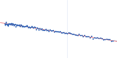
Synchrotron SAXS data from solutions of K352D Pcs60p in 50 mM Hepes, 150 mM NaCl, 0.5 mM TCEP, pH 7.5 were collected on the EMBL P12 beam line at PETRA III (DESY, Hamburg, Germany) using a Pilatus 6M detector at a sample-detector distance of 3 m and at a wavelength of λ = 0.124 nm (I(s) vs s, where s = 4πsinθ/λ, and 2θ is the scattering angle). One solute concentration of 0.50 mg/ml was measured at 10°C. 38 successive 0.095 second frames were collected. The data were normalized to the intensity of the transmitted beam and radially averaged; the scattering of the solvent-blank was subtracted.
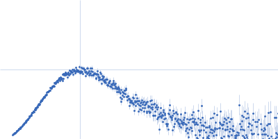
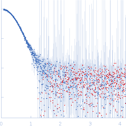
Oxalate--CoA ligase (K352D) (Pcs60-K352D)
Mol. type   Protein
Organism   Saccharomyces cerevisiae (strain ATCC 204508 / S288c)
Olig. state   Dimer
Mon. MW   60.5 kDa
 
UniProt   P38137 (1-543)
Sequence   FASTA