Form factor determination of biological molecules with X-ray free electron laser small-angle scattering (XFEL-SAS).

Blanchet CE, Round A, Mertens HDT, Ayyer K, Graewert M, Awel S, Franke D, Dörner K, Bajt S, Bean R, Custódio TF, de Wijn R, Juncheng E, Henkel A, Gruzinov A, Jeffries CM, Kim Y, Kirkwood H, Kloos M, Knoška J, Koliyadu J, Letrun R, Löw C, Makroczyova J, Mall A, Meijers R, Pena Murillo GE, Oberthür D, Round E, Seuring C, Sikorski M, Vagovic P, Valerio J, Wollweber T, Zhuang Y, Schulz J, Haas H, Chapman HN, Mancuso AP, Svergun D, Commun Biol 6(1):1057 (2023) Europe PMC

SASDQ29 – Thyroglobulin - reservoir sample storage with liquid jet delivery measured using XFEL

Thyroglobulin
MWexperimental 300 kDa
MWexpected 303 kDa
log I(s) 1.31×10⁻² 1.31×10⁻³ 1.31×10⁻⁴ 1.31×10⁻⁵
Thyroglobulin small angle scattering data  s, nm-1
ln I(s)
Thyroglobulin Guinier plot ln 1.31×10⁻² Rg: 7.0 nm 0 (7.0 nm)-2 s2
(sRg)2I(s)/I(0)
Thyroglobulin Kratky plot 1.104 0 3 sRg

Data validation


There are no models related to this curve.
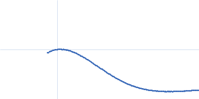
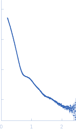
Scattering data from solutions of Thyroglobulin - reservoir sample storage with liquid jet delivery measured using XFEL in phosphate buffered saline, pH 7.4 were collected using a AGIPD-1Mpix detector at a sample-detector distance of 3 m and at a wavelength of λ = 0.13 nm (I(s) vs s, where s = 4πsinθ/λ, and 2θ is the scattering angle). . The data were normalized to the intensity of the transmitted beam and radially averaged; the scattering of the solvent-blank was subtracted.

Thyroglobulin solutions were measured on the European XFEL using a "reservoir" to inject the sample into an in-vacuum liquid jet delivery system. There is no definable Guinier region.

Thyroglobulin
Mol. type   Protein
Organism   Bos taurus
Olig. state   Monomer
Mon. MW   303.2 kDa
 
UniProt   P01267 (1-2769)
Sequence   FASTA