Npl3 functions in mRNP assembly by recruitment of mRNP components to the transcription site and their transfer onto the mRNA.

Keil P, Wulf A, Kachariya N, Reuscher S, Hühn K, Silbern I, Altmüller J, Keller M, Stehle R, Zarnack K, Sattler M, Urlaub H, Sträßer K, Nucleic Acids Res (2022) Europe PMC

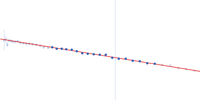
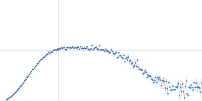
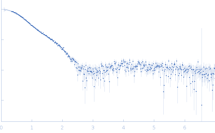
SASDQT5 – NPL3 protein RNA recognition motifs 1 and 2 (RRM1,2) wild-type

Serine/arginine (SR)-type shuttling mRNA binding protein NPL3
MWI(0) 18 kDa
MWexpected 18 kDa
VPorod 24 nm3
log I(s) 4.78×10⁻² 4.78×10⁻³ 4.78×10⁻⁴ 4.78×10⁻⁵
Serine/arginine (SR)-type shuttling mRNA binding protein NPL3 small angle scattering data  s, nm-1
ln I(s)
Serine/arginine (SR)-type shuttling mRNA binding protein NPL3 Guinier plot ln 4.78×10⁻² Rg: 2 nm 0 (2 nm)-2 s2
(sRg)2I(s)/I(0)
Serine/arginine (SR)-type shuttling mRNA binding protein NPL3 Kratky plot 1.104 0 3 sRg
p(r)
Serine/arginine (SR)-type shuttling mRNA binding protein NPL3 pair distance distribution function Rg: 2.1 nm 0 Dmax: 6.8 nm

Data validation


There are no models related to this curve.

SAXS data from solutions of the NPL3 protein RNA recognition motif domains 1 and 2 (RRM1,2) in 20 mM NaPO4, 50 mM NaCl, 1 mM DTT, pH 6.5 were collected using a Rigaku bioSAXS-1000 instrument at the Technische Universität München (TUM; Garching, Germany) equipped with a Pilatus 100K detector at a sample-detector distance of 0.5 m and at a wavelength of λ = 0.155 nm (I(s) vs s, where s = 4πsinθ/λ, and 2θ is the scattering angle). One solute concentration of 4.90 mg/ml was measured at 25°C. Eight successive 900 second frames were collected. The data were normalized to the intensity of the transmitted beam and radially averaged; the scattering of the solvent-blank was subtracted.

Sample was dialyzed in same buffer before the measurements.

Serine/arginine (SR)-type shuttling mRNA binding protein NPL3 (Tandem RRMs)
Mol. type   Protein
Organism   Saccharomyces cerevisiae (strain ATCC 204508 / S288c)
Olig. state   Monomer
Mon. MW   18.2 kDa
 
UniProt   Q01560 (120-280)
Sequence   FASTA