|
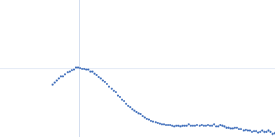
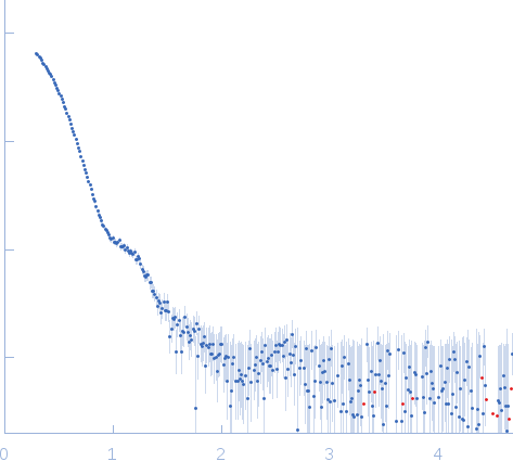
Synchrotron SAXS
data from solutions of
Pyruvate decarboxylase (PDC) from Z. mobilis
in
100 mM Sodium Citrate, 17% Glycerol, 22.5% PEG 1500, pH 6
were collected
on the
EMBL X33 beam line
at the DORIS III, DESY storage ring
(Hamburg, Germany)
using a 1D Gas detector detector
(I(s) vs s, where s = 4πsinθ/λ, and 2θ is the scattering angle).
.
The data were normalized to the intensity of the transmitted beam and radially averaged; the scattering of the solvent-blank was subtracted.
The low angle data collected at lower concentration were merged with the highest concentration high angle data to yield the final composite scattering curve.
Wavelength = UNKNOWN. Cell temperature = UNKNOWN. Storage temperature = UNKNOWN. Sample detector distance = UNKNOWN. X-ray Exposure time = UNKNOWN. Number of frames = UNKNOWN. Concentration min = UNKNOWN. Concentration max = UNKNOWN
|
|
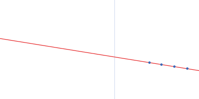
 s, nm-1