Death-Associated Protein Kinase Activity Is Regulated by Coupled Calcium/Calmodulin Binding to Two Distinct Sites.

Simon B, Huart AS, Temmerman K, Vahokoski J, Mertens HD, Komadina D, Hoffmann JE, Yumerefendi H, Svergun DI, Kursula P, Schultz C, McCarthy AA, Hart DJ, Wilmanns M, Structure 24(6):851-61 (2016) Europe PMC

SASDB52 – Death associated protein kinase (wild-type)

Death associated protein kinase wild-type
MWexperimental 41 kDa
MWexpected 37 kDa
VPorod 101 nm3
log I(s) 7.95×10² 7.95×101 7.95×100 7.95×10⁻¹
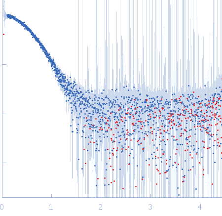
Death associated protein kinase wild-type small angle scattering data  s, nm-1
ln I(s)
Death associated protein kinase wild-type Guinier plot ln 7.95×10² Rg: 2.7 nm 0 (2.7 nm)-2 s2
(sRg)2I(s)/I(0)
Death associated protein kinase wild-type Kratky plot 1.104 0 3 sRg
p(r)
Death associated protein kinase wild-type pair distance distribution function Rg: 2.6 nm 0 Dmax: 8.1 nm

Data validation


Fits and models


log I(s)
 s, nm-1
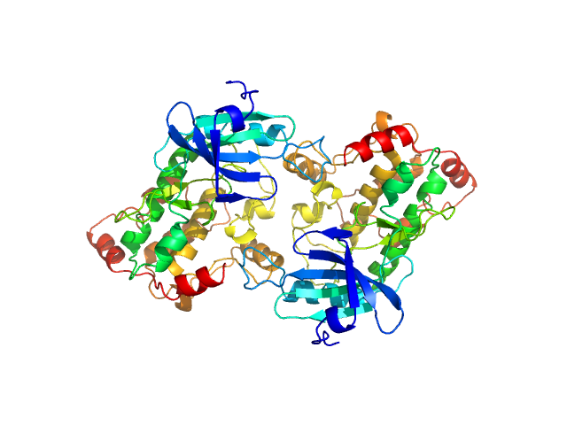
Death associated protein kinase wild-type NONE model

Synchrotron SAXS data from solutions of Death associated protein kinase (wild-type) in 50 mM HEPES 250 mM NaCl 5mM CaCl2 0.25 mM TCEP 5% (v/v) glycerol, pH 7.5 were collected on the EMBL P12 beam line at the PETRA III storage ring (DESY; Hamburg, Germany) using a Pilatus 2M detector at a sample-detector distance of 3.1 m and at a wavelength of λ = 0.12 nm (I(s) vs s, where s = 4πsinθ/λ, and 2θ is the scattering angle). One solute concentration of 2.00 mg/ml was measured at 10°C. 20 successive 0.050 second frames were collected. The data were normalized to the intensity of the transmitted beam and radially averaged; the scattering of the solvent-blank was subtracted.

Characterisation of the oligomeric equilibrium of human DAPK2 protein in solution.

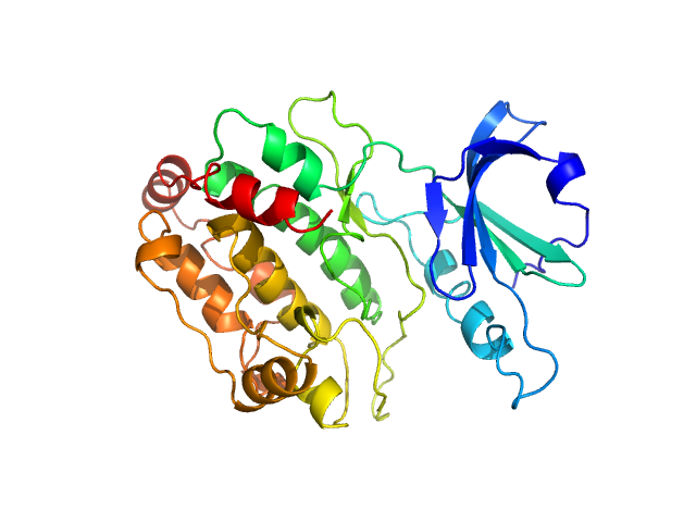
Death associated protein kinase wild-type (hDAPK2 WT)
Mol. type   Protein
Organism   Homo sapiens
Olig. state   Other
Mon. MW   37.3 kDa
Sequence   FASTA
 
PDB ID   2A2A