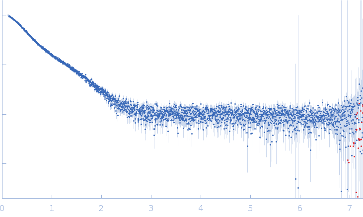
Synchrotron SAXS data from solutions of myotilin 220-452 (wild type) in 20 mM HEPES, 150 mM NaCl, 5 % glycerol, 1 mM DTT, pH 7.4 were collected on the EMBL P12 beam line at PETRA III (DESY, Hamburg, Germany) using a Pilatus 6M detector at a sample-detector distance of 3 m and at a wavelength of λ = 0.124 nm (I(s) vs s, where s = 4πsinθ/λ, and 2θ is the scattering angle). The data displayed here show the results obtained from one solute concentration of 5.09 mg/ml of a dilution series that was measured at 20°C. The data were normalized to the intensity of the transmitted beam and radially averaged; the scattering of the solvent-blank was subtracted.
Our previous structural studies suggest full-length myotilin is monomeric under our experimental conditions. To investigate molecular determinants of dimeriaation SAXS was used on the truncated construct Ig1Ig2 220-452. We observed a concentration-dependent increase in the average molecular mass. Accordingly, the P(r) function showed a transition from a typical distribution for an extended two-domain protein, with a significant peak at approximately 2.5 nm and a minor at 5.0 nm, to a distribution with increased frequencies of the 5.0 nm peak and a new minor signal at in the region 8-10 nm. SAXS data measured from the concentration series are made available as additional files in the full-entry zip-archive.
 s, nm-1