Engineering the NK1 Fragment of Hepatocyte Growth Factor/Scatter Factor as a MET Receptor Antagonist

Youles M, Holmes O, Petoukhov M, Nessen M, Stivala S, Svergun D, Gherardi E, Journal of Molecular Biology 377(3):616-622 (2008) DOI

SASDLZ5 – Hepatocyte growth factor/Hepatocyte growth factor receptor (NK1-Met567) complex

Hepatocyte growth factor
Hepatocyte growth factor receptor
MWexperimental 210 kDa
MWexpected 161 kDa
VPorod 290 nm3
log I(s) 8.45×10² 8.45×101 8.45×100 8.45×10⁻¹
Hepatocyte growth factor Hepatocyte growth factor receptor small angle scattering data  s, nm-1
ln I(s)
Hepatocyte growth factor Hepatocyte growth factor receptor Guinier plot ln 8.46×10² Rg: 5.3 nm 0 (5.3 nm)-2 s2
(sRg)2I(s)/I(0)
Hepatocyte growth factor Hepatocyte growth factor receptor Kratky plot 1.104 0 3 sRg
p(r)
Hepatocyte growth factor Hepatocyte growth factor receptor pair distance distribution function Rg: 5.5 nm 0 Dmax: 20 nm

Data validation


Fits and models


log I(s)
 s, nm-1
Hepatocyte growth factor Hepatocyte growth factor receptor MONSA model

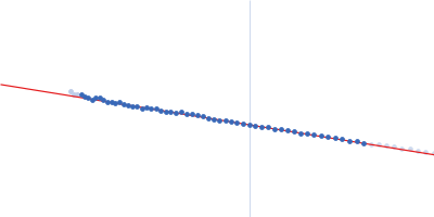
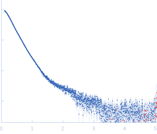
Synchrotron SAXS data from solutions of the NK1-Met567 complex in PBS containing EDTA, surfactant P20 and 5 M NaCl, pH 6, were collected on the EMBL X33 beam line at DORIS III (DESY, Hamburg, Germany) using a MAR 345 Image Plate detector at a wavelength of λ = 0.15 nm (I(s) vs s, where s = 4πsinθ/λ, and 2θ is the scattering angle). One solute concentration of 5.00 mg/ml was measured. The data were normalized to the intensity of the transmitted beam and radially averaged; the scattering of the solvent-blank was subtracted.

X-ray exposure time: UNKNOWN. Experimental temperature: UNKNOWN. Sample-to-detector distance: UNKNOWN.

Tags: X33
Hepatocyte growth factor (NK1)
Mol. type   Protein
Organism   Homo sapiens
Olig. state   Dimer
Mon. MW   19.7 kDa
 
UniProt   P14210 (38-209)
Sequence   FASTA
 
Hepatocyte growth factor receptor (Met567)
Mol. type   Protein
Organism   Homo sapiens
Olig. state   Dimer
Mon. MW   61.0 kDa
 
UniProt   P08581 (25-562)
Sequence   FASTA