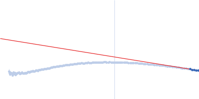
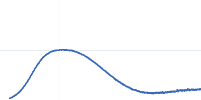
Solution Structure of Poly(ethylene) Glycol-Conjugated Hemoglobin Revealed by Small-Angle X-Ray Scattering: Implications for a New Oxygen Therapeutic

Svergun D, Ekström F, Vandegriff K, Malavalli A, Baker D, Nilsson C, Winslow R, Biophysical Journal 94(1):173-181 (2008) DOI

SASDNN2 – Experimental SAXS data for hemoglobin conjucted with six-seven copies of PEG dimer (Hb2) at concentration c = 21 mg/ml

Human hemoglobin conjugated with six-seven copies of 5-kDa PEG
MWexperimental 62 kDa
MWexpected 62 kDa
log I(s) 1.07×101 1.07×100 1.07×10⁻¹ 1.07×10⁻²
Human hemoglobin conjugated with six-seven copies of 5-kDa PEG small angle scattering data  s, nm-1
ln I(s)
Human hemoglobin conjugated with six-seven copies of 5-kDa PEG Guinier plot ln 1.07×101 Rg: 3.0 nm 0 (3.0 nm)-2 s2
(sRg)2I(s)/I(0)
Human hemoglobin conjugated with six-seven copies of 5-kDa PEG Kratky plot 1.104 0 3 sRg

Data validation


There are no models related to this curve.
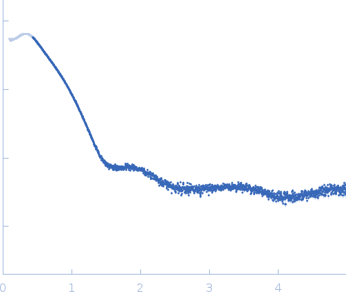
Synchrotron SAXS data from solutions of Experimental SAXS data for hemoglobin conjucted with six-seven copies of PEG dimer (Hb2) at concentration c = 21 mg/ml in Ringer's lactate solution, pH 6.5 were collected on the EMBL X33 beam line at the DORIS III, DESY storage ring (Hamburg, Germany) using a MAR 345 Image Plate detector at a sample-detector distance of 2.5 m and at a wavelength of λ = 0.15 nm (I(s) vs s, where s = 4πsinθ/λ, and 2θ is the scattering angle). One solute concentration of 21.00 mg/ml was measured. Three successive 60 second frames were collected. The data were normalized to the intensity of the transmitted beam and radially averaged; the scattering of the solvent-blank was subtracted.

Cell temperature = UNKNOWN. Storage temperature = UNKNOWN

Tags: X33
Human hemoglobin conjugated with six-seven copies of 5-kDa PEG (hb5)
Mol. type   Protein
Organism   Homo sapiens
Olig. state   Dimer
Mon. MW   30.8 kDa
Sequence   FASTA