|
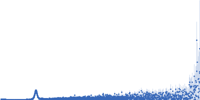
Synchrotron SAXS data from solutions of cationic liposomes containing DOTMA:DOPE (2:1) with the negatively charged messenger RNA (mRNA) (raito 0.65:1) in 10 mM HEPES, 5 mM NaCl, 0.1 mM EDTA, pH 7.4 were collected on the EMBL P12 beam line at PETRA III (DESY; Hamburg, Germany) using a Pilatus 6M detector at a sample-detector distance of 3 m and at a wavelength of λ = 0.154 nm (I(s) vs s, where s = 4πsinθ/λ, and 2θ is the scattering angle). The samples were fractionated by an asymmetrical flow field-flow fractionation (AF4) system (PN AF2000 MT). A semi-preparative frit-inlet AF4 (Fl-AF4) channel (shoulder width 50 mm, tip width 5 mm, tip-to-tip length 277 mm) was equipped with a polyethersulfone PES membrane with 10 kDa molecular weight cut-off and a Mylar spacer of 350 µm height. The temperature of the autosampler was set to a constant temperature of 4 °C. The system was hyphenated to a UV detector (PN 3211) and a MALS detector (PN 3621, 21 angles). The UV absorbance wavelength was set to 260 nm. The MALS detector angles were normalized with respect to 90° using a fractionated 62 nm PS-NP size standard. The MALS was directly connected to the SAXS flow capillary (set to 20°C) using a minimal tubing length with an ID of 250 µm.
The selected peak fittings (top to bottom) show the consecutive elution of the mRNA liposomes through the AF4 fractionation at 31, 38, 46, 55 and 63 minutes.
R-Dotma: https://pubchem.ncbi.nlm.nih.gov/compound/118978115
DOPE: https://pubchem.ncbi.nlm.nih.gov/compound/1_2-Dioleoyl-sn-glycero-3-phosphoethanolamine
|
|
 s, nm-1
![mRNA -- proprietary sequence (R)-N,N,N-trimethyl-2-3-dioleyloxy-1-propanaminium chloride 1,2-dioleoyl-sn-glycero-3-phosphoethanolamine OTHER [STATIC IMAGE] model](/media//pdb_file/SASDSH7_fit1_model1.png "Static model image")