|
|
|
|
|
| Sample: |
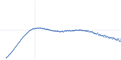
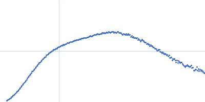
Human immunoglobulin gamma 2 (IgG2) SAP1.3 F(ab')2 - heavy chain C227S dimer, 52 kDa protein
Human immunoglobulin gamma 2 (IgG2) SAP1.3 F(ab')2 - kappa light chain C214S dimer, 48 kDa Homo sapiens protein
|
| Buffer: |
50 mM HEPES, 150 mM KCl, pH: 7.5
|
| Experiment: |
SAXS
data collected at BM29, ESRF on 2022 Jun 8
|
Structure-guided disulfide engineering restricts antibody conformation to elicit TNFR agonism.
Nat Commun 16(1):3495 (2025)
Elliott IG, Fisher H, Chan HTC, Inzhelevskaya T, Mockridge CI, Penfold CA, Duriez PJ, Orr CM, Herniman J, Müller KTJ, Essex JW, Cragg MS, Tews I
|
| RgGuinier |
3.9 |
nm |
| Dmax |
13.7 |
nm |
| VolumePorod |
135 |
nm3 |
|
|
|
|
|
|
|
| Sample: |
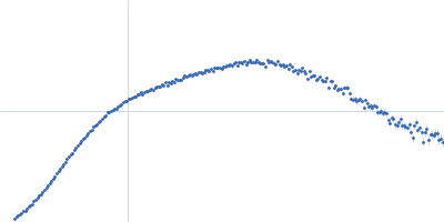
Human immunoglobulin gamma 2 (IgG2) SAP1.3 F(ab')2 - heavy chain C226S/C227S dimer, 52 kDa Homo sapiens protein
Human immunoglobulin gamma 2 (IgG2) SAP1.3 F(ab')2 - kappa light chain dimer, 48 kDa Homo sapiens protein
|
| Buffer: |
50 mM HEPES, 150 mM KCl, pH: 7.5
|
| Experiment: |
SAXS
data collected at BM29, ESRF on 2022 Jun 8
|
Structure-guided disulfide engineering restricts antibody conformation to elicit TNFR agonism.
Nat Commun 16(1):3495 (2025)
Elliott IG, Fisher H, Chan HTC, Inzhelevskaya T, Mockridge CI, Penfold CA, Duriez PJ, Orr CM, Herniman J, Müller KTJ, Essex JW, Cragg MS, Tews I
|
| RgGuinier |
4.2 |
nm |
| Dmax |
15.1 |
nm |
| VolumePorod |
140 |
nm3 |
|
|
|
|
|
|
|
| Sample: |
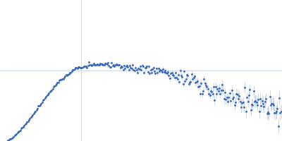
Human immunoglobulin gamma 2 (IgG2) SAP1.3 F(ab')2 - kappa light chain dimer, 48 kDa Homo sapiens protein
Human immunoglobulin gamma 2 (IgG2) SAP1.3 F(ab')2 - heavy chain dimer, 52 kDa Homo sapiens protein
|
| Buffer: |
50 mM HEPES, 150 mM KCl, pH: 7.5
|
| Experiment: |
SAXS
data collected at BM29, ESRF on 2022 Jun 8
|
Structure-guided disulfide engineering restricts antibody conformation to elicit TNFR agonism.
Nat Commun 16(1):3495 (2025)
Elliott IG, Fisher H, Chan HTC, Inzhelevskaya T, Mockridge CI, Penfold CA, Duriez PJ, Orr CM, Herniman J, Müller KTJ, Essex JW, Cragg MS, Tews I
|
| RgGuinier |
3.8 |
nm |
| Dmax |
12.8 |
nm |
| VolumePorod |
126 |
nm3 |
|
|
|
|
|
|
|
| Sample: |
Human immunoglobulin gamma 1 (IgG1) SAP1.3 F(ab')2 - heavy chain dimer, 51 kDa Homo sapiens protein
Human immunoglobulin gamma 1 (IgG1) SAP1.3 F(ab')2 - kappa light chain dimer, 48 kDa Homo sapiens protein
|
| Buffer: |
50 mM HEPES, 150 mM KCl, pH: 7.5
|
| Experiment: |
SAXS
data collected at BM29, ESRF on 2022 Jun 8
|
Structure-guided disulfide engineering restricts antibody conformation to elicit TNFR agonism.
Nat Commun 16(1):3495 (2025)
Elliott IG, Fisher H, Chan HTC, Inzhelevskaya T, Mockridge CI, Penfold CA, Duriez PJ, Orr CM, Herniman J, Müller KTJ, Essex JW, Cragg MS, Tews I
|
| RgGuinier |
4.5 |
nm |
| Dmax |
16.3 |
nm |
| VolumePorod |
139 |
nm3 |
|
|
|
|
|
|
|
| Sample: |
Human immunoglobulin gamma 1 (IgG1) ChiLob 7/4 F(ab')2 - heavy chain dimer, 51 kDa Homo sapiens protein
Human immunoglobulin gamma 1 (IgG1) ChiLob 7/4 F(ab')2 - kappa light chain dimer, 47 kDa Homo sapiens protein
|
| Buffer: |
50 mM HEPES, 150 mM KCl, pH: 7.5
|
| Experiment: |
SAXS
data collected at BM29, ESRF on 2022 Apr 12
|
Structure-guided disulfide engineering restricts antibody conformation to elicit TNFR agonism.
Nat Commun 16(1):3495 (2025)
Elliott IG, Fisher H, Chan HTC, Inzhelevskaya T, Mockridge CI, Penfold CA, Duriez PJ, Orr CM, Herniman J, Müller KTJ, Essex JW, Cragg MS, Tews I
|
| RgGuinier |
4.6 |
nm |
| Dmax |
17.4 |
nm |
| VolumePorod |
133 |
nm3 |
|
|
|
|
|
|
|
| Sample: |
Human immunoglobulin gamma 2 (IgG2) ChiLob 7/4 F(ab')2 - heavy chain dimer, 52 kDa Homo sapiens protein
Human immunoglobulin gamma 2 (IgG2) ChiLob 7/4 F(ab')2 - kappa light chain dimer, 47 kDa Homo sapiens protein
|
| Buffer: |
50 mM HEPES, 150 mM KCl, pH: 7.5
|
| Experiment: |
SAXS
data collected at BM29, ESRF on 2022 Jun 8
|
Structure-guided disulfide engineering restricts antibody conformation to elicit TNFR agonism.
Nat Commun 16(1):3495 (2025)
Elliott IG, Fisher H, Chan HTC, Inzhelevskaya T, Mockridge CI, Penfold CA, Duriez PJ, Orr CM, Herniman J, Müller KTJ, Essex JW, Cragg MS, Tews I
|
| RgGuinier |
3.9 |
nm |
| Dmax |
13.7 |
nm |
| VolumePorod |
123 |
nm3 |
|
|
|
|
|
|
|
| Sample: |
Human immunoglobulin gamma 2 (IgG2) ChiLob 7/4 F(ab')2 - heavy chain C224S/C225S dimer, 52 kDa Homo sapiens protein
Human immunoglobulin gamma 2 (IgG2) ChiLob 7/4 F(ab')2 - kappa light chain dimer, 47 kDa Homo sapiens protein
|
| Buffer: |
50 mM HEPES, 150 mM KCl, pH: 7.5
|
| Experiment: |
SAXS
data collected at BM29, ESRF on 2022 Apr 12
|
Structure-guided disulfide engineering restricts antibody conformation to elicit TNFR agonism.
Nat Commun 16(1):3495 (2025)
Elliott IG, Fisher H, Chan HTC, Inzhelevskaya T, Mockridge CI, Penfold CA, Duriez PJ, Orr CM, Herniman J, Müller KTJ, Essex JW, Cragg MS, Tews I
|
| RgGuinier |
4.2 |
nm |
| Dmax |
14.8 |
nm |
| VolumePorod |
127 |
nm3 |
|
|
|
|
|
|
|
| Sample: |
Human immunoglobulin gamma 2 (IgG2) ChiLob 7/4 F(ab')2 - kappa light chain C214S dimer, 47 kDa Homo sapiens protein
Human immunoglobulin gamma 2 (IgG2) ChiLob 7/4 F(ab')2 - heavy chain C225S dimer, 52 kDa Homo sapiens protein
|
| Buffer: |
50 mM HEPES, 150 mM KCl, pH: 7.5
|
| Experiment: |
SAXS
data collected at BM29, ESRF on 2022 Apr 12
|
Structure-guided disulfide engineering restricts antibody conformation to elicit TNFR agonism.
Nat Commun 16(1):3495 (2025)
Elliott IG, Fisher H, Chan HTC, Inzhelevskaya T, Mockridge CI, Penfold CA, Duriez PJ, Orr CM, Herniman J, Müller KTJ, Essex JW, Cragg MS, Tews I
|
| RgGuinier |
4.0 |
nm |
| Dmax |
14.1 |
nm |
| VolumePorod |
125 |
nm3 |
|
|
|
|
|
|
|
| Sample: |
Human immunoglobulin gamma 2 (IgG2) ChiLob 7/4 F(ab')2 - kappa light chain C214S dimer, 47 kDa Homo sapiens protein
Human immunoglobulin gamma 2 (IgG2) ChiLob 7/4 F(ab')2 - heavy chain C224S dimer, 52 kDa Homo sapiens protein
|
| Buffer: |
50 mM HEPES, 150 mM KCl, pH: 7.5
|
| Experiment: |
SAXS
data collected at BM29, ESRF on 2022 Apr 12
|
Structure-guided disulfide engineering restricts antibody conformation to elicit TNFR agonism.
Nat Commun 16(1):3495 (2025)
Elliott IG, Fisher H, Chan HTC, Inzhelevskaya T, Mockridge CI, Penfold CA, Duriez PJ, Orr CM, Herniman J, Müller KTJ, Essex JW, Cragg MS, Tews I
|
| RgGuinier |
3.9 |
nm |
| Dmax |
13.9 |
nm |
| VolumePorod |
126 |
nm3 |
|
|
|
|
|
|
|
| Sample: |
Human immunoglobulin gamma 2 (IgG2) ChiLob 7/4 - heavy chain dimer, 99 kDa Homo sapiens protein
Human immunoglobulin ChiLob 7/4 - kappa chain dimer, 47 kDa Homo sapiens protein
|
| Buffer: |
50 mM HEPES, 150 mM KCl, pH: 7.5
|
| Experiment: |
SAXS
data collected at BM29, ESRF on 2024 Sep 28
|
Structure-guided disulfide engineering restricts antibody conformation to elicit TNFR agonism.
Nat Commun 16(1):3495 (2025)
Elliott IG, Fisher H, Chan HTC, Inzhelevskaya T, Mockridge CI, Penfold CA, Duriez PJ, Orr CM, Herniman J, Müller KTJ, Essex JW, Cragg MS, Tews I
|
| RgGuinier |
4.9 |
nm |
| Dmax |
16.0 |
nm |
| VolumePorod |
202 |
nm3 |
|
|
 SAP1.3 F(ab')2 - heavy chain C227SHuman immunoglobulin gamma 2 (IgG2) SAP1.3 F(ab')2 - kappa light chain C214S experimental SAS data")
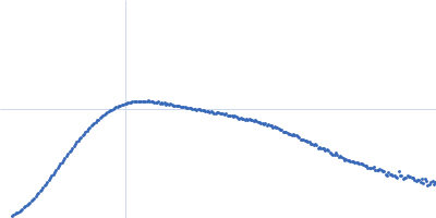
 SAP1.3 F(ab')2 - heavy chain C226S/C227SHuman immunoglobulin gamma 2 (IgG2) SAP1.3 F(ab')2 - kappa light chain experimental SAS data")
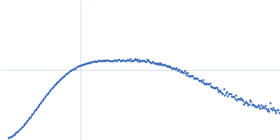
 SAP1.3 F(ab')2 - kappa light chainHuman immunoglobulin gamma 2 (IgG2) SAP1.3 F(ab')2 - heavy chain experimental SAS data")
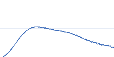
 SAP1.3 F(ab')2 - heavy chainHuman immunoglobulin gamma 1 (IgG1) SAP1.3 F(ab')2 - kappa light chain experimental SAS data")
 ChiLob 7/4 F(ab')2 - heavy chainHuman immunoglobulin gamma 1 (IgG1) ChiLob 7/4 F(ab')2 - kappa light chain experimental SAS data")
 ChiLob 7/4 F(ab')2 - heavy chainHuman immunoglobulin gamma 2 (IgG2) ChiLob 7/4 F(ab')2 - kappa light chain experimental SAS data")
 ChiLob 7/4 F(ab')2 - heavy chain C224S/C225SHuman immunoglobulin gamma 2 (IgG2) ChiLob 7/4 F(ab')2 - kappa light chain experimental SAS data")
 ChiLob 7/4 F(ab')2 - heavy chain C224S/C225S mutant Rg histogram")
 ChiLob 7/4 F(ab')2 - kappa light chain C214SHuman immunoglobulin gamma 2 (IgG2) ChiLob 7/4 F(ab')2 - heavy chain C225S experimental SAS data")
 ChiLob 7/4 F(ab')2 - heavy chain C225S kappa chain C214S mutant Rg histogram")
 ChiLob 7/4 F(ab')2 - kappa light chain C214SHuman immunoglobulin gamma 2 (IgG2) ChiLob 7/4 F(ab')2 - heavy chain C224S experimental SAS data")
 ChiLob 7/4 F(ab')2 - heavy chain C224S kappa chain C214S mutant Rg histogram")
 ChiLob 7/4 - heavy chainHuman immunoglobulin ChiLob 7/4 - kappa chain experimental SAS data")