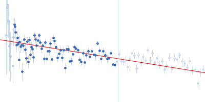
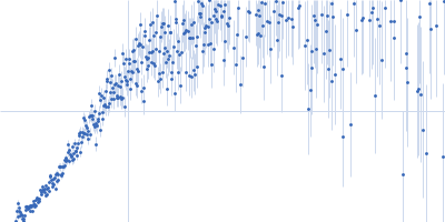
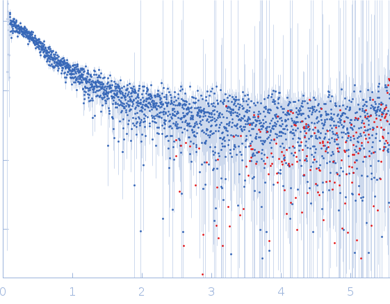
Synchrotron SAXS data from solutions of Peroxisome Proliferator-Activated Receptor Gamma Activation Function-1 Domain (AF1) in 10 mM sodium phosphate, pH 7.4, containing 50 mM NaCl in 10 mM sodium phosphate, 50 mM NaCl, pH 7.4 were collected on the ID7A1 BioSAXS / HP-Bio Beamline beam line at the Cornell High Energy Synchrotron Source (CHESS) storage ring (Ithaca, NY, USA) using a Eiger 4M detector at a sample-detector distance of 1.7 m and at a wavelength of λ = 0.110659 nm (I(s) vs s, where s = 4πsinθ/λ, and 2θ is the scattering angle). A 100 µL sample (10 mg/ml) was injected onto a Superdex 200 Increase 5/150 GL column and eluted isocratically at 0.30 mL/min at 4 °C, with 1 s SAXS exposures collected throughout the SEC run.The SEC-SAXS series was processed in BioXTAS RAW 2.3.0. Evolving factor analysis (EFA) was performed over frames 350–560, identifying one significant scattering component. The component corresponding to the main SEC elution peak was selected over frames 387–560, while the adjacent shoulder was excluded to avoid combining scattering from distinct populations. The resulting EFA-derived scattering profile was used for Guinier and Bayesian indirect Fourier transform (BIFT) analyses to determine the radius of gyration and pair-distance distribution.

 s, nm-1