Szameit K,
Berg K,
Kruspe S,
Valentini E
Magbanua E,
Kwiatkowski M,
Chauvot de Beauchêne I,
Krichel B,
Schamoni K,
Uetrecht C,
Svergun DI,
Schlüter H,
Zacharias M,
Hahn U,
RNA Biol
13(10):973-987
(2016)
Europe PMC
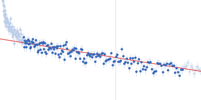
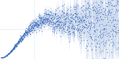
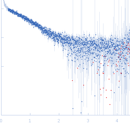
Synchrotron SAXS
data from solutions of
Aptamer AIR-3A 2'FU
in
water, pH 7.5
were collected
on the
EMBL P12 beam line
at the PETRA III storage ring
(DESY; Hamburg, Germany)
using a Pilatus 2M detector
at a sample-detector distance of 3.1 m and
at a wavelength of λ = 0.12 nm
(I(s) vs s, where s = 4πsinθ/λ, and 2θ is the scattering angle).
One solute concentration of 0.30 mg/ml was measured
at 10°C.
20 successive
0.050 second frames were collected.
The data were normalized to the intensity of the transmitted beam and radially averaged; the scattering of the solvent-blank was subtracted.
 s, nm-1