Structure and target interaction of a G-quadruplex RNA-aptamer.

Szameit K, Berg K, Kruspe S, Valentini E Magbanua E, Kwiatkowski M, Chauvot de Beauchêne I, Krichel B, Schamoni K, Uetrecht C, Svergun DI, Schlüter H, Zacharias M, Hahn U, RNA Biol 13(10):973-987 (2016) Europe PMC

SASDAZ7 – IL-6R AIR-3A 2:4 complex

AIR-3A
Interleukin-6 receptor subunit alpha
MWexperimental 150 kDa
MWexpected 107 kDa
VPorod 150 nm3
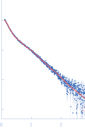
log I(s) 3.62×10³ 3.62×10² 3.62×101 3.62×100
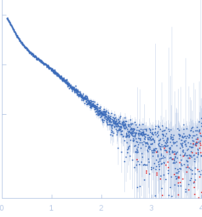
AIR-3A Interleukin-6 receptor subunit alpha small angle scattering data  s, nm-1
ln I(s)
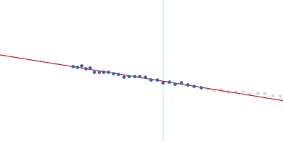
AIR-3A Interleukin-6 receptor subunit alpha Guinier plot ln 3.63×10³ Rg: 6.7 nm 0 (6.7 nm)-2 s2
(sRg)2I(s)/I(0)
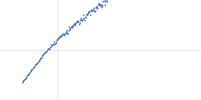
AIR-3A Interleukin-6 receptor subunit alpha Kratky plot 1.104 0 3 sRg
p(r)
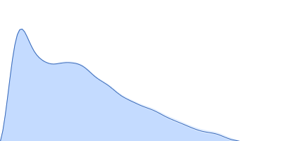
AIR-3A Interleukin-6 receptor subunit alpha pair distance distribution function Rg: 6.6 nm 0 Dmax: 25 nm

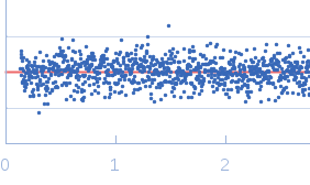
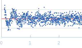
Data validation


Fits and models


log I(s)
 s, nm-1
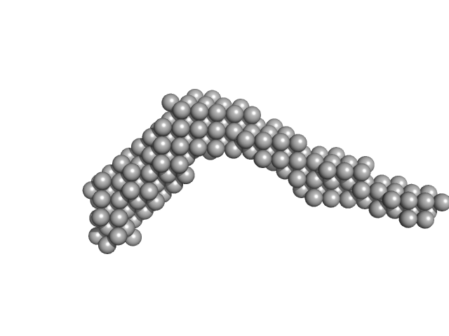
AIR-3A Interleukin-6 receptor subunit alpha DAMMIF model

log I(s)
 s, nm-1
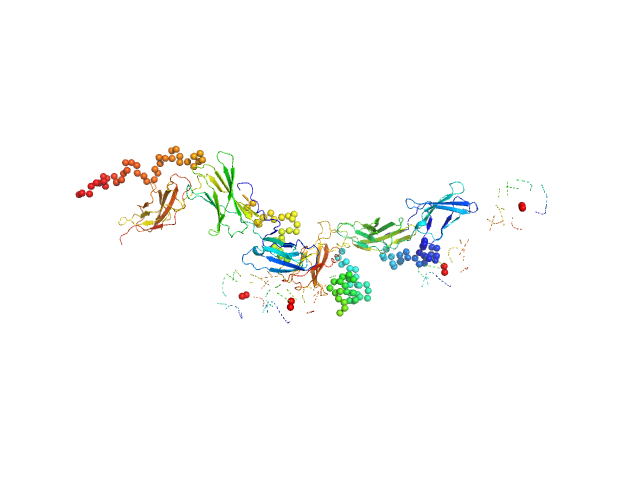
AIR-3A Interleukin-6 receptor subunit alpha CORAL model

Synchrotron SAXS data from solutions of IL-6R AIR-3A 2:4 complex in water, pH 7.5 were collected on the B21 beam line at the Diamond Light Source storage ring (Didcot, UK) using a Pilatus 2M detector at a sample-detector distance of 3.9 m and (I(s) vs s, where s = 4πsinθ/λ, and 2θ is the scattering angle). One solute concentration of 0.20 mg/ml was measured. The data were normalized to the intensity of the transmitted beam and radially averaged; the scattering of the solvent-blank was subtracted.

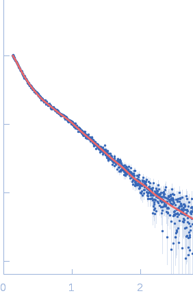
The CORAL model fits the experimental curve with a Chi-squared-value of 1.43. The model appears slightly smaller probably because of the presence of free RNA dimers in solution.

AIR-3A
Mol. type   RNA
Olig. state   Monomers
Mon. MW   6.4 kDa
Sequence   FASTA
 
Interleukin-6 receptor subunit alpha (IL-6R)
Mol. type   Protein
Organism   Homo sapiens
Olig. state   Dimer
Mon. MW   41.0 kDa
 
UniProt   P08887
Sequence   FASTA
 
PDB ID   1N26