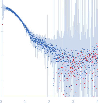
X-ray synchrotron radiation scattering data were measured from solutions of Human Calumenin in 25 mM NaHEPES, 25 mM NaCl, 2.5 mM CaCl2 pH 7.5 buffer using size exclusion chromatography-SAXS (HPLC SEC-SAXS) on the B21 beam line of the DIAMOND storage ring (Oxfordshire, UK). The sample load concentration was 13 mg/mL. The scattering data, I(s) vs s (s = 4π sin θ/λ, where 2θ is the scattering angle) were collected through the corresponding SEC protein elution peak (41 data frames) as well as from the SEC column running buffer, and radially averaged to produce 1D I(s) vs s scattering profiles. The buffer scattering contributions were subtracted from each sample frame, which were then scaled and averaged to produce the SAXS profile displayed in this entry. Data were recorded using a Pilatus 2M detector at an experimental temperature of 20 degrees centigrade.
Wavelength = UNKNOWN. Cell temperature = UNKNOWN. X-ray Exposure time = UNKNOWN. Concentration = UNKNOWN
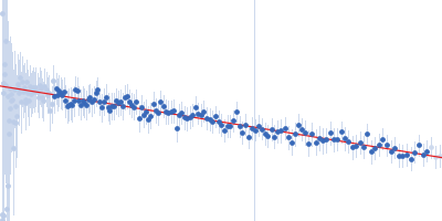
 s, nm-1
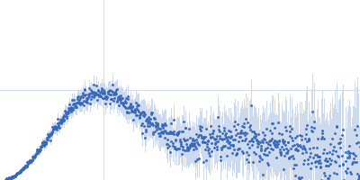