A novel Porphyromonas gingivalis enzyme: An atypical dipeptidyl peptidase III with an ARM repeat domain.

Hromić-Jahjefendić A, Jajčanin Jozić N, Kazazić S, Grabar Branilović M, Karačić Z, Schrittwieser JH, Das KMP, Tomin M, Oberer M, Gruber K, Abramić M, Tomić S, PLoS One 12(11):e0188915 (2017) Europe PMC

SASDC58 – Dipeptidyl peptidase III: pgDPP3_FL

Peptidase, M49 family
MWI(0) 101 kDa
MWexpected 102 kDa
VPorod 116 nm3
log I(s) 7.02×101 7.02×100 7.02×10⁻¹ 7.02×10⁻²
Peptidase, M49 family small angle scattering data  s, nm-1
ln I(s)
Peptidase, M49 family Guinier plot ln 7.02×101 Rg: 3 nm 0 (3 nm)-2 s2
(sRg)2I(s)/I(0)
Peptidase, M49 family Kratky plot 1.104 0 3 sRg
p(r)
Peptidase, M49 family pair distance distribution function Rg: 3.1 nm 0 Dmax: 11.3 nm

Data validation


Fits and models


log I(s)
 s, nm-1
Peptidase, M49 family DAMMIN model

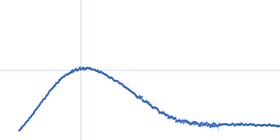
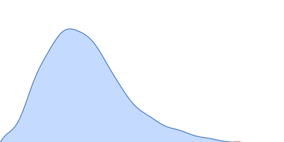
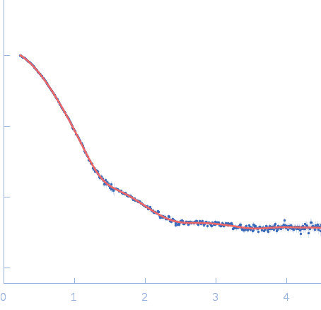
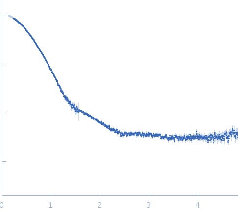
Synchrotron SAXS data from solutions of Dipeptidyl peptidase III: pgDPP3_FL in 50mM Tris,100mM NaCl, pH 8, were collected on the BM29 beam line at the ESRF storage ring (Grenoble, France) using a Pilatus 1M detector at a sample-detector distance of 2.5 m (I(s) vs s, where s = 4πsinθ/λ, and 2θ is the scattering angle). Solute concentrations ranging between 1.0 and 8.8 mg/ml were measured . Eight successive frames were collected. The data were normalized to the intensity of the transmitted beam and radially averaged; the scattering of the solvent-blank was subtracted and the different curves were scaled for protein concentration. The low angle data collected at lower concentration were merged with the highest concentration high angle data to yield the final composite scattering curve.

X-ray Exposure time = UNKNOWN

Peptidase, M49 family (PgDPP3)
Mol. type   Protein
Organism   Porphyromonas gingivalis
Olig. state   Monomer
Mon. MW   101.7 kDa
 
UniProt   Q7MX92
Sequence   FASTA