Solution structure of the 128 kDa enzyme I dimer from Escherichia coli and its 146 kDa complex with HPr using residual dipolar couplings and small- and wide-angle X-ray scattering.

Schwieters CD, Suh JY, Grishaev A, Ghirlando R, Takayama Y, Clore GM, J Am Chem Soc 132(37):13026-45 (2010) Europe PMC

SASDCN3 – Phosphoenolpyruvate-protein phosphotransferase

Phosphoenolpyruvate-protein phosphotransferase
MWexperimental 128 kDa
MWexpected 127 kDa
VPorod 189 nm3
log I(s) 2.90×10⁻² 2.90×10⁻³ 2.90×10⁻⁴ 2.90×10⁻⁵
Phosphoenolpyruvate-protein phosphotransferase small angle scattering data  s, nm-1
ln I(s)
Phosphoenolpyruvate-protein phosphotransferase Guinier plot ln 2.90×10⁻² Rg: 4.1 nm 0 (4.1 nm)-2 s2
(sRg)2I(s)/I(0)
Phosphoenolpyruvate-protein phosphotransferase Kratky plot 1.104 0 3 sRg
p(r)
Phosphoenolpyruvate-protein phosphotransferase pair distance distribution function Rg: 4.2 nm 0 Dmax: 14.8 nm

Data validation


Fits and models


log I(s)
 s, nm-1
Phosphoenolpyruvate-protein phosphotransferase PDB (PROTEIN DATA BANK) model

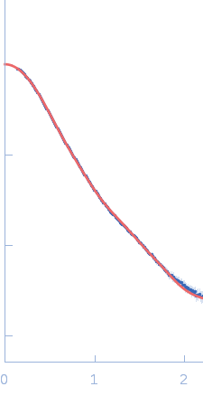
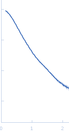
Synchrotron SAXS data from a solution of Phosphoenolpyruvate-protein phosphotransferase in 20 mM TRIS buffer, pH 7.4, 100 mM NaCl, 10 mM DTT, 4 mM MgCl2, 1 mM EDTA, and 1 tablet of protease inhibitor cocktail (SigmaFAST S8830) were acquired at the beamline 12ID-C at the Advanced Photon Source (APS, Argonne, IL, USA). One solute concentration of 5 mg/ml (corresponding to ∼40 μM dimer) was measured at 25°C using a Gold CCD detector at a sample-detector distance of 4 m and at a wavelength of λ = 0.06199 nm (s = 4π sin θ/λ, where 2θ is the scattering angle). 20 successive 0.25 second frames were collected. To prevent radiation damage, volumes of 150 μL of samples and buffers were oscillated during data collection using a flow-though setup. Individual data frames were masked, corrected for the detector sensitivity, radially integrated and normalized by the corresponding incident beam intensities. The final 1D scattering profiles and their uncertainties were calculated as means and standard deviations over the 20 individual frames. The buffer data were then subtracted from the scattering profiles.

Storage temperature = UNKNOWN

Phosphoenolpyruvate-protein phosphotransferase (Enzyme I)
Mol. type   Protein
Organism   Escherichia coli
Olig. state   Dimer
Mon. MW   63.6 kDa
 
UniProt   A0A037YGN3 (1-575)
Sequence   FASTA
 
PDB ID   2KX9