Dmax unknown – experimental data range validation not possible.
There are no models related to this curve.
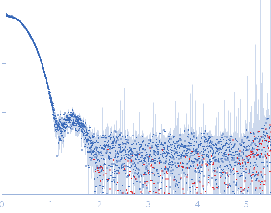
Synchrotron SAXS data from solutions of glucose isomerase in 100 mM HEPES, 1 mM MgCl2, pH 7 were collected at the EMBL-P12 beam line at the PETRA III storage ring (Hamburg, Germany) using an Eiger 4M detector at a sample-detector distance of 1.6 m and at a wavelength of λ = 0.124 nm (I(s) vs s, where s = 4πsinθ/λ and 2θ is the scattering angle). One solute concentration of 4.07 mg/ml was measured at 20°C. A single 1.3 millisecond frame was collected. The data were normalized to the intensity of the transmitted beam and radially averaged; the scattering of the solvent-blank was subtracted and the data were scaled to the protein concentration.
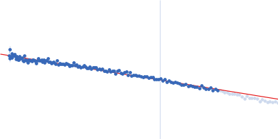
Only one concentration was measured. There might be some repulsive interactions as the guinier region is not linear and the Rg and volume are smaller than expected.
 s, nm-1
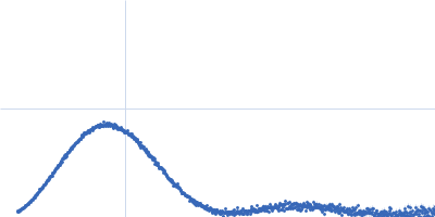