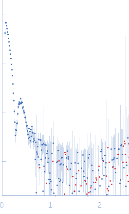
Dmax unknown – experimental data range validation not possible.
There are no models related to this curve.
Synchrotron SAXS data from solutions of Bacteriophage phi-X174 - Empty Capsid in 0.06 M NH4Cl2, 0.09 M NaCl, 0.1 M KCl, 1 mM MgS04, 1 mM CaCl2, 0.1 M Tris-HCl, pH 7.4 were collected on the BioSAXS G1 beam line at the Cornell High Energy Synchrotron Source (CHESS; Ithaca, NY, USA) using a Pilatus Pilatus 200K detector at a sample-detector distance of 2.1 m and at a wavelength of λ = 0.1109 nm (I(s) vs s, where s = 4πsinθ/λ and 2θ is the scattering angle). One solute concentration of 0.10 mg/ml phage was measured at 33°C. 10 successive 10 second frames were collected. The data were normalized to the intensity of the transmitted beam and radially averaged; the scattering of the solvent-blank was subtracted.
Note: Due to the size of the capsids, the Rg and I(0) obtained from Guinier analysis should be interpreted as approximated estimates only.
 s, nm-1
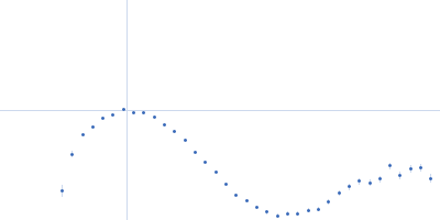