Structures of insect Imp-L2 suggest an alternative strategy for regulating the bioavailability of insulin-like hormones.

Roed NK, Viola CM, Kristensen O, Schluckebier G, Norrman M Sajid W, Wade JD, Andersen AS, Kristensen C, Ganderton TR, Turkenburg JP, De Meyts P, Brzozowski AM, Nat Commun 9(1):3860 (2018) Europe PMC

SASDDS8 – Neural/ectodermal development factor IMP-L2

Neural/ectodermal development factor IMP-L2
MWexperimental 63 kDa
MWexpected 60 kDa
VPorod 93 nm3
log I(s) 4.09×101 4.09×100 4.09×10⁻¹ 4.09×10⁻²
Neural/ectodermal development factor IMP-L2 small angle scattering data  s, nm-1
ln I(s)
Neural/ectodermal development factor IMP-L2 Guinier plot ln 4.09×101 Rg: 3.1 nm 0 (3.1 nm)-2 s2
(sRg)2I(s)/I(0)
Neural/ectodermal development factor IMP-L2 Kratky plot 1.104 0 3 sRg
Dmax: 12 nm

Data validation


There are no models related to this curve.

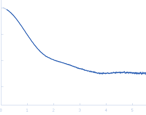
Synchrotron SAXS data from solutions of neural/ectodermal development factor IMP-L2 in phosphate buffered saline, pH 7.4 were collected on the ID14-3 beam line at the ESRF (Grenoble, France) using a Pilatus 1M detector at a sample-detector distance of 2.4 m and at a wavelength of λ = 0.093 nm (l(s) vs s, where s = 4πsinθ/λ, and 2θ is the scattering angle). Solute concentrations ranging between 3.2 and 12.8 mg/ml were measured at 25°C. 10 successive 10 second frames were collected. The data were normalized to the intensity of the transmitted beam and radially averaged; the scattering of the solvent-blank was subtracted. The low angle data collected at lower concentration were merged with the highest concentration high angle data to yield the final composite scattering curve.
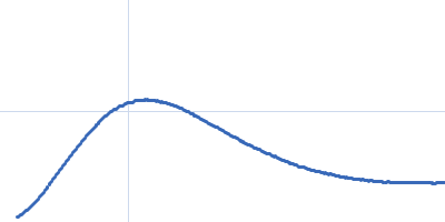
Neural/ectodermal development factor IMP-L2 (Imp-L2 IBP)
Mol. type   Protein
Organism   Drosophila melanogaster
Olig. state   Dimer
Mon. MW   29.9 kDa
 
UniProt   Q09024
Sequence   FASTA