|
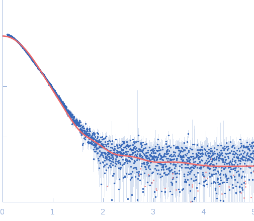
Synchrotron SAXS data from solutions of the MoCh construct of MICAL1 in 50 mM sodium phosphate buffer, 5 % glycerol, 100 mM NaCl, 1 mM EDTA, 1 mM DTT, pH 7.5 were collected on the EMBL P12 beam line at the PETRA III storage ring (Hamburg, Germany) using a Pilatus 2M detector at a sample-detector distance of 3 m and at a wavelength of λ = 0.124 nm (l(s) vs s, where s = 4πsinθ/λ, and 2θ is the scattering angle). One solute concentration of 4.47 mg/ml was measured at 15°C. 20 successive 0.050 second frames were collected. The data were normalized to the intensity of the transmitted beam and radially averaged; the scattering of the solvent-blank was subtracted.
|
|
![[F-actin]-monooxygenase MICAL1 (MoCh) small angle scattering data](/media/intensities_files/scattering_plots/SASDDU9_dat_img.png) s, nm-1
s, nm-1
![[F-actin]-monooxygenase MICAL1 (MoCh) PDB (PROTEIN DATA BANK) model](/media/pdb_file/images/SASDDU9_fit1_model1_img.png "Load 3D view")
![[F-actin]-monooxygenase MICAL1 (MoCh) Guinier plot](/media//intensities_files/scattering_plots/SASDDU9_guinier_img.png)
![[F-actin]-monooxygenase MICAL1 (MoCh) Kratky plot](/media/intensities_files/scattering_plots/SASDDU9_kratky_img.png)
![[F-actin]-monooxygenase MICAL1 (MoCh) pair distance distribution function](/media/p_of_R_files/pofr_images/SASDDU9_pofr_img.png)