Synchrotron SAXS measurements (l(s) vs s, where s = 4πsinθ/λ, and 2θ is the scattering angle; λ = 0.11 nm) were carried out at the beam line X12SA of the Swiss Light Source (Paul Scherrer Institut, Villigen, Switzerland). Quartz capillaries (inner diameter 1 mm) were mounted in a sample holder and filled with buffer (20 mM HEPES, 150 mM NaCl, 5 mM MgCl2, 5 % w/v glycerol, pH 7.5). The sample holder was cooled to 10 °C for all measurements. Scattering of the buffer was determined at 20 positions along the capillary (spacing 0.5 mm), exposing each position with 11.2 keV photons for 0.5 s using 200 successive frames. Each position was measured 10 times and scattering intensities, after radial averaging to produce 1D-scattering patterns, were averaged. Scattered X-rays were detected by a Pilatus 2M detector (Dectris, Baden-Dättwil, Switzerland) at the end of an evacuated flight tube, 2.13m from the sample position. Buffer was replaced by the protein sample without moving the sample holder or the capillaries. Lights in the experimental hutch were switched off and after 5 minutes, scattering of the protein-containing solutions was measured at the same positions using the procedure described above. Due to poor behaviour of this sample, molecular weight and Rg estimates from the Guinier region need to be treated with caution. This sample is mostly provided to illustrate the differences that occur upon nucleotide binding of mPAC. Solute concentrations ranging between 4.7 and 7.5 mg/ml were measured and the concentration series data extrapolated to infinite dilution generating the final SAXS profile displayed in this entry.
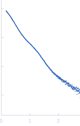 s, nm-1