|
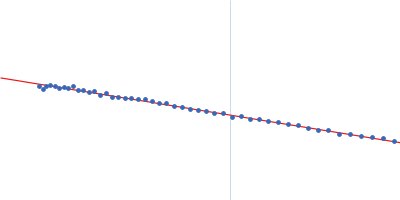
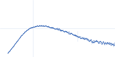
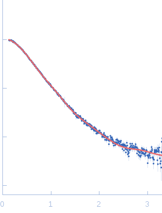
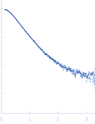
Synchrotron SAXS data from solutions of Interleukin-18 receptor 1, IL-18Rα-ECD in 10mM HEPES, 150mM NaCl, 3% glycerol, pH 7.2 were measured using size-exclusion chromatography SAXS (SEC-SAXS) on the 12.3.1 (SIBYLS) beam line at the Advanced Light Source (ALS; Berkeley, CA, USA) using a using a MAR 165 CCD detector at a sample-detector distance of 1.5 m and at a wavelength of λ = 0.103 nm (l(s) vs s, where s = 4πsinθ/λ, and 2θ is the scattering angle). The data were collected as 600 successive 3 second frames through the SEC elution profile. Sample-elution peak data were normalized to the intensity of the transmitted beam and radially averaged; the scattering of the solvent-blank was subtracted and the individual subtracted data sets were scaled and averaged to generate the scattering profile displayed in this entry.
SEC-SAXS was performed at 20°C using the following parameters: Column: Schodex kw-803 ; Flow rate: 0.5 mL/min; Total acquisition time: 30min; Sample injection concentration: 10 mg/mL; Injection volume: 50 μL.
|
|
 s, nm-1