Cooperative DNA Binding of the Plasmid Partitioning Protein TubR from the Bacillus cereus pXO1 Plasmid.

Hayashi I, Oda T, Sato M, Fuchigami S, J Mol Biol (2018) Europe PMC

SASDEH3 – TubR protein of the pXO1-like plasmid pBc10987 from B. cereus (Bc-TubR) bound to S48 DNA (Bc-TubR : S48 DNA complex)

S48 DNA strand 1
S48 DNA strand 2
TubR of the pXO1-like plasmid pBc10987 from B. cereus (Bc-TubR)
MWexperimental 249 kDa
MWexpected 179 kDa
VPorod 305 nm3
log I(s) 7.95×10⁻² 7.95×10⁻³ 7.95×10⁻⁴ 7.95×10⁻⁵
S48 DNA strand 1 S48 DNA strand 2 TubR of the pXO1-like plasmid pBc10987 from B. cereus (Bc-TubR) small angle scattering data  s, nm-1
ln I(s)
S48 DNA strand 1 S48 DNA strand 2 TubR of the pXO1-like plasmid pBc10987 from B. cereus (Bc-TubR) Guinier plot ln 7.95×10⁻² Rg: 6.1 nm 0 (6.1 nm)-2 s2
(sRg)2I(s)/I(0)
S48 DNA strand 1 S48 DNA strand 2 TubR of the pXO1-like plasmid pBc10987 from B. cereus (Bc-TubR) Kratky plot 1.104 0 3 sRg
p(r)
S48 DNA strand 1 S48 DNA strand 2 TubR of the pXO1-like plasmid pBc10987 from B. cereus (Bc-TubR) pair distance distribution function Rg: 6.6 nm 0 Dmax: 23 nm

Data validation


Fits and models


log I(s)
 s, nm-1
S48 DNA strand 1 S48 DNA strand 2 TubR of the pXO1-like plasmid pBc10987 from B. cereus (Bc-TubR) MOLECULAR DYNAMICS FRAME model

log I(s)
 s, nm-1
S48 DNA strand 1 S48 DNA strand 2 TubR of the pXO1-like plasmid pBc10987 from B. cereus (Bc-TubR) DAMFILT model

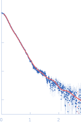
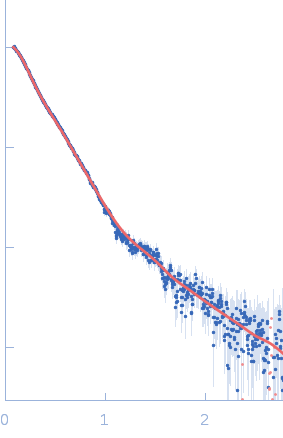
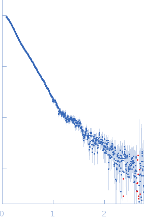
Synchrotron SAXS data from a solution of the Bc-TubR:S48 DNA complex in 0.1 M NaCl, 10 mM Tris, pH 8 were collected on the BL-10C beam line at the Photon Factory (PF), High Energy Accelerator Research Organization (KEK) storage ring (Tsukuba, Japan) using a Pilatus3 2M detector at a sample-detector distance of 2.0 m and at a wavelength of λ = 0.15 nm (l(s) vs s, where s = 4πsinθ/λ, and 2θ is the scattering angle). Data were collected for a 20 s X-ray exposure time at a sample temperature of 8°C. The data were normalized to the intensity of the transmitted beam and radially averaged; the scattering of the solvent-blank was subtracted.

The molecular weight (MW) of the Bc-TubR : S48 DNA complex was estimated from the macromolecular volume using the method of Fischer et al. (J. Appl. Crystallogr. 43, 101–109, 2010; SAXMow program). However, the MW estimate is likely not an accurate value because the sample is a complex of protein and DNA. The theoretical SAXS curve of the molecular dynamics model was calculated as the mixture of the two models (one model contains four TubR dimers plus DNA, another model contains five TubR dimers plus DNA). For the mixed model, the fractions of each are 0.4 and 0.6, respectively. A low resolution dummy atom model was built by DAMMIF. Ten independently built models were generated, spatially aligned and then bead-occupancy and volume corrected using DAMAVER to generate an averaged spatial representation of the complex (damfilt). The the fit to the SAXS data from an individual dummy atom model from the cohort of models used to obtain the average representation is shown, specifically that with the smallest chi-square value. Sample concentration: UNKNOWN

S48 DNA strand 1
Mol. type   DNA
Olig. state   Monomer
Mon. MW   21.3 kDa
Sequence   FASTA
 
S48 DNA strand 2
Mol. type   DNA
Olig. state   Monomer
Mon. MW   21.3 kDa
Sequence   FASTA
 
TubR of the pXO1-like plasmid pBc10987 from B. cereus (Bc-TubR) (Bc-TubR)
Mol. type   Protein
Olig. state   Decamer
Mon. MW   13.7 kDa
 
UniProt   B7JTH0 (1-116)
Sequence   FASTA