Structural Organization and Dynamics of Homodimeric Cytohesin Family Arf GTPase Exchange Factors in Solution and on Membranes.

Das S, Malaby AW, Nawrotek A, Zhang W Zeghouf M, Maslen S, Skehel M, Chakravarthy S, Irving TC, Bilsel O, Cherfils J, Lambright DG, Structure (2019) Europe PMC

SASDEV9 – Cytohesin-2; ARF nucleotide-binding site opener, ARNO truncation mutant

Cytohesin-2; ARNO truncation mutant
MWexperimental 42 kDa
MWexpected 40 kDa
VPorod 63 nm3
log I(s) 3.04×10⁻² 3.04×10⁻³ 3.04×10⁻⁴ 3.04×10⁻⁵
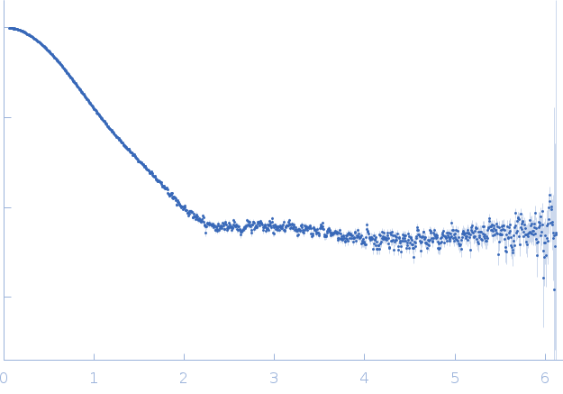
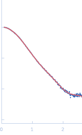
Cytohesin-2; ARNO truncation mutant small angle scattering data  s, nm-1
ln I(s)
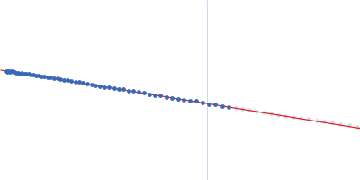
Cytohesin-2; ARNO truncation mutant Guinier plot ln 3.04×10⁻² Rg: 2.7 nm 0 (2.7 nm)-2 s2
(sRg)2I(s)/I(0)
Cytohesin-2; ARNO truncation mutant Kratky plot 1.104 0 3 sRg
p(r)
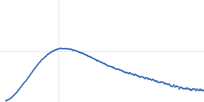
Cytohesin-2; ARNO truncation mutant pair distance distribution function Rg: 2.8 nm 0 Dmax: 9.9 nm

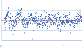
Data validation


Fits and models


log I(s)
 s, nm-1
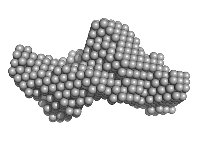
Cytohesin-2; ARNO truncation mutant DAMMIN model

Synchrotron SAXS data from solutions ofthe cytohesin-2; ARF nucleotide-binding site opener, ARNO truncation mutant in 300 mM NaCl, 2 mM 2-mercaptoethanol and 30 mM Tris-HCl, pH 7.5 were collected on the SWING beam line at SOLEIL (Saint-Aubin, France) using a CCD AVIEX PCCD170170 detector at a sample-detector distance of 1.8 m and at a wavelength of λ = 0.1033 nm (l(s) vs s, where s = 4πsinθ/λ, and 2θ is the scattering angle). One solute concentration of 8.00 mg/ml was measured at 15°C. One 1.5 second frame was collected. The data were normalized to the intensity of the transmitted beam and radially averaged; the scattering of the solvent-blank was subtracted.

Cytohesin-2; ARNO truncation mutant (ARNO-CC)
Mol. type   Protein
Organism   Homo sapiens
Olig. state   Monomer
Mon. MW   39.9 kDa
 
UniProt   Q99418 (58-400)
Sequence   FASTA