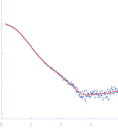
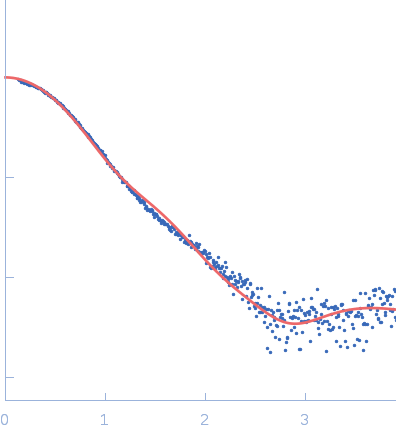
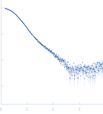
| MWI(0) | 36 | kDa |
| MWexpected | 42 | kDa |
| VPorod | 45 | nm3 |
|
log I(s)
5.49×10²
5.49×101
5.49×100
5.49×10⁻¹
|
 s, nm-1
|
|
|
|

|
|

|
|
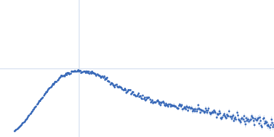
SAXS data from solutions of gamma-crystallin S, disulfide-linked dimer, in 20 mM sodium phosphate, pH 7 were collected on the Bruker Nanostar II instrument at the Australian Nuclear Science and Technology Organisation/Australian Centre for Neutron Scattering (ANSTO/ACNS, Sydney, Australia) using a multiwire Bruker Hi-Star detector at a sample-detector distance of 0.7 m and at a wavelength of λ = 0.1541 nm (l(s) vs s, where s = 4πsinθ/λ, and 2θ is the scattering angle). One solute concentration of 6.00 mg/ml was measured at 25°C. Four successive 3600 second frames were collected. The data were normalized to the intensity of the transmitted beam and radially averaged; the scattering of the solvent-blank was subtracted.
|
|
|||||||||||||||||||||||||||||||||