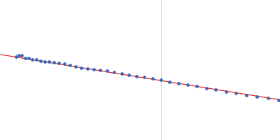
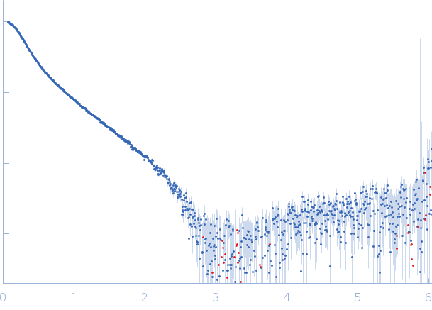
Synchrotron SAXS data from solutions of the R16-24 del45-55 human dystrophin fragment in NaP 20 mM, NaCl 300 mM, EDTA 1 mM, glycerol 2%, pH 7.5 were collected using size-exclusion chromatography SAXS (SEC-SAXS) on the SWING beam line at SOLEIL (Saint-Aubin, France) using a CCD AVIEX PCCD170170 detector at a sample-detector distance of 1.8 m and at a wavelength of λ = 0.1033 nm (l(s) vs s, where s = 4πsinθ/λ, and 2θ is the scattering angle). The data were normalized to the intensity of the transmitted beam and radially averaged; the scattering of the solvent-blank was subtracted.
SEC-SAXS was performed at 20°C using the following parameters: Column: BioSEC5-500Å (4.6 mm id * 300 mm); Flow rate: 0.2 mL/min; Sample injection concentration: ~8 mg/mL; Injection volume: 50μL. The data were collected through the SEC peak of the protein as a series of 21 x 1.5 second exposures.
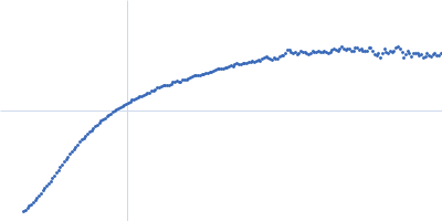
The experimental molecular weight was determined from the volume of correlation, Vc.
 s, nm-1