Dmax unknown – experimental data range validation not possible.
There are no models related to this curve.
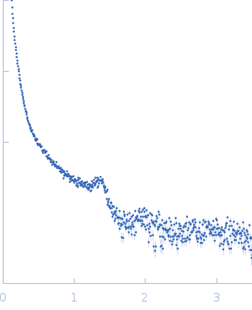
Synchrotron SAXS data from solutions of DNA-binding protein HU-alpha bound to 80 base-pair DNA in 10 mM sodium acetate, 100 mM NaCl, pH 4.5 were collected on the 12.3.1 (SIBYLS) beam line at the Advanced Light Source (ALS) storage ring (Berkeley, CA, USA) using a Pilatus3 X 2M detector at a sample-detector distance of 1.5 m and at a wavelength of λ = 0.103 nm (I(s) vs s, where s = 4πsinθ/λ, and 2θ is the scattering angle). 300 successive 3 second frames were collected at 10°C. The data were normalized to the intensity of the transmitted beam and radially averaged; the scattering of the solvent-blank was subtracted.
SAXS profile corresponds to a protein-DNA complex between bacterial nucleoid associated protein HUalpha and 80bp DNA. Under these conditions, the complex forms lamellar structures visible as Bragg Diffraction peaks.
 s, nm-1

