Protease-associated import systems are widespread in Gram-negative bacteria.

Grinter R, Leung PM, Wijeyewickrema LC, Littler D, Beckham S, Pike RN, Walker D, Greening C, Lithgow T, PLoS Genet 15(10):e1008435 (2019) Europe PMC

SASDFB6 – The periplasmically localised protease PqqL from Escherichia coli

Zinc protease PqqL
MWexperimental 113 kDa
MWexpected 102 kDa
VPorod 207 nm3
log I(s) 1.36×10⁻¹ 1.36×10⁻² 1.36×10⁻³ 1.36×10⁻⁴
Zinc protease PqqL small angle scattering data  s, nm-1
ln I(s)
Zinc protease PqqL Guinier plot ln 1.37×10⁻¹ Rg: 4.0 nm 0 (4.0 nm)-2 s2
(sRg)2I(s)/I(0)
Zinc protease PqqL Kratky plot 1.104 0 3 sRg
p(r)
Zinc protease PqqL pair distance distribution function Rg: 4.1 nm 0 Dmax: 13.7 nm

Data validation


Fits and models


log I(s)
 s, nm-1
Zinc protease PqqL DAMMIF model

log I(s)
 s, nm-1
Zinc protease PqqL PDB (PROTEIN DATA BANK) model

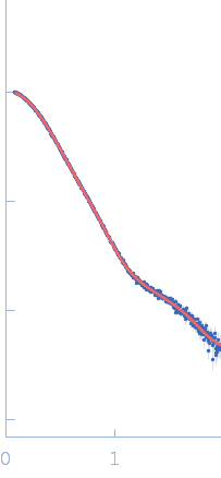
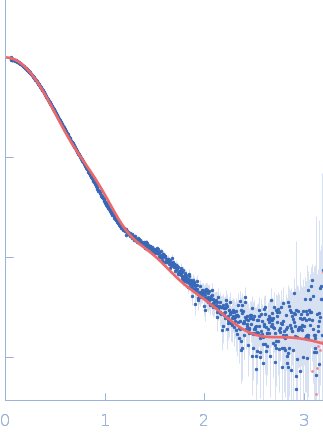
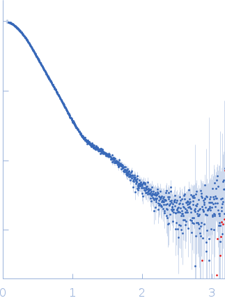
Synchrotron SAXS data from solutions of periplasmically localised protease (PqqL) from E. coli in 20 mM Tris HCl, 150 nM NaCl, 0.02 % NaN3, 5% glycerol, pH 7.8 were collected on the SAXS/WAXS beam line at the Australian Synchrotron (Melbourne, Australia) using a Pilatus 1M detector at a sample-detector distance of 1.4 m and at a wavelength of λ = 0.10322 nm (I(s) vs s, where s = 4πsinθ/λ, and 2θ is the scattering angle). In-line size-exclusion chromatography (SEC) SAXS was employed using a co-flow apparatus. The SEC parameters were as follows: A 100 μl sample at 8 mg/ml was injected onto a GE Superdex 200 Increase 5/150 column at 20°C. 10 successive 1 second frames were collected through the SEC elution peak. The data were normalized to the intensity of the transmitted beam and radially averaged; the scattering of the solvent-blank was subtracted.

Zinc protease PqqL
Mol. type   Protein
Organism   Escherichia coli
Olig. state   Monomer
Mon. MW   101.7 kDa
 
UniProt   P31828 (27-931)
Sequence   FASTA
 
PDB ID   6OFS