Dmax unknown – experimental data range validation not possible.
There are no models related to this curve.
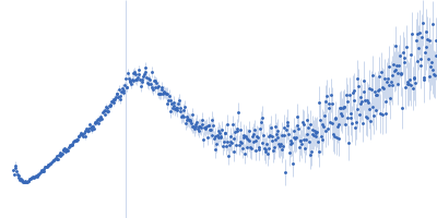
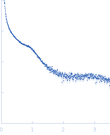
Synchrotron SAXS data from solutions of DNA-binding protein HU-alpha bound to 80 base-pair DNA in 10mM Bis-Tris, 50 mM NaCl, pH 6.5 were collected on the 12.3.1 (SIBYLS) beam line at the Advanced Light Source (ALS) storage ring (Berkeley, CA, USA) using a Pilatus3 X 2M detector at a sample-detector distance of 1.5 m and at a wavelength of λ = 0.103 nm (I(s) vs s, where s = 4πsinθ/λ, and 2θ is the scattering angle). 300 successive 3 second frames were collected at 10°C. The data were normalized to the intensity of the transmitted beam and radially averaged; the scattering of the solvent-blank was subtracted.
SAXS profile corresponds to a protein-DNA complex between bacterial nucleoid associated protein HUalpha and 80bp DNA. Under these conditions, the complex forms lamellar structures visible as Bragg Diffraction peaks.
 s, nm-1